Download
1 / 1
10 likes | 148 Views
How to get from the EPR spectrum of a tyrosyl radical in a protein to the identity of the Tyr residue that hosts the radical. input. q. Or download from: http://www.biophysj.org/content/vol87/issue1/images/ data/582/DC1/svistunenko_online_materials.zip

E N D
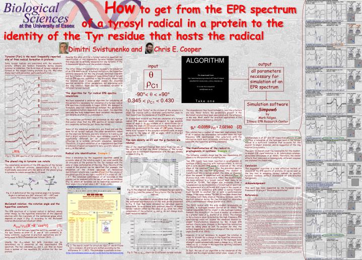
How to get from the EPR spectrum of a tyrosyl radical in a protein to the identity of the Tyr residue that hosts the radical input q Or download from: http://www.biophysj.org/content/vol87/issue1/images/ data/582/DC1/svistunenko_online_materials.zip Or e-mail me a request at: svist@essex.ac.uk rC1 ALGORITHM P.Denitrificans CcO+H2O2 (MacMillan et al. 1999); Horse metMb+H2O2 (Miki et al. 1989); Mouse TA3 cells RNR (Sahlin et al. 1987); E.coli RNR (Hoganson and Babcock 1992); Ovine PGHS, “wide doublet” (Kulmacz et al. 1990); Ovine PGHS, “narrow singlet” (Dorlet et al. 2002); Soybean metLb+H2O2 (Davies and Puppo 1992); S.typhimurium RNR (Allard et al. 1996); A.thaliana photosystem II YD (Svistunenko et al. 2002); Human metHb+H2O2 (Svistunenko et al. 2002); Bovine catalase+peroxyacetic acid (Ivancich et al. 1996). output all parameters necessary for simulation of an EPR spectrum -90°< q < +90° 0.345 < rC1 < 0.430 Simulation software Simpow6 by Mark Nilges, Illinois EPR Research Center T a k e o n e 20 G at the University of Essex Tyrosine (Tyr) is the most frequently reported site of free radical formation in proteins Some tyrosyl radicals are associated with the enzymatic activity. Others are formed transiently during enzyme turnover or inhibition. The third class of tyrosyl radicals are formed on the oxygen binding proteins (e.g. Hb, Mb) when those react with peroxides, particularly with H2O2. Knowing the value of q for a tyrosyl radical is important for identification of the responsible tyrosine residue, because this angle can be directly measured for any tyrosine, if the 3-dimensional structure of the protein is known. The ‘other things’ (the parameters) necessary for simulation of an EPR spectrum of a tyrosyl radical are the hyperfine splitting constants for the ring protons, individual linewidth and the g-factors.An analysis of many simulations of tyrosyl radical EPR spectra, published in the literature, shows that the rotation angle q alone cannot explain the observed spectral variability. Other parameters, not associated with the ring rotation, must also be considered variables in different tyrosyl radicals. The algorithm for Tyr radical EPR spectra simulations We have suggested a universal algorithm for calculating all the parameters necessary for simulation of a tyrosyl radical EPR spectrum (Svistunenko & Cooper 2004). We managed to formulate empirical relationships between the parameters not considered before dependent. As a result, the algorithm uses only two truly independent input variables: the unpaired spin density on atom C1 (rC1) and angle q. The simulations performed and presented on the right of this poster show that the EPR spectra of tyrosyl radicals are completely defined by just these two values. Some of the simulation parameters are fixed and are the same for all tyrosyl radicals. The other parameters, linked via the empirical formulae, are very sensitive to q and rC1, so that a slight variation in these input variables results in a significantly different set of simulation parameters, effecting in a notably different simulation spectrum. Therefore, if a good simulation of an experimental spectrum is achieved, it would correspond to very accurate values of q and rC1 in the radical. Radical site identification. Example 1. Once a simulation by the suggested algorithm yields an accurate value of the rotation angle q, we could search the 3D structure of the protein for the Tyr residues with the same or close conformation of the ring. Thus, the site of the radical in the protein can be identified. For example, the simulation of the X-band spectrum of Paracoccus denitrificans cytochrome c oxidase (Panel 2 in the column of simulated spectra on the right) results in a q-values of -38 ° ± 2 °. The rotational conformation of all tyrosines in this enzyme can be analyzed with the use of our on-line database (Fig. 3), where all tyrosines can be ranked by closeness of their angle q to a target value (-38 ° in our case). Fig. 3 shows that Tyr167 on the A-chain of the enzyme is a single Tyr residue with a rotational conformation close to that found from the simulation of the EPR spectrum. It is important to point out that any simulation of a tyrosyl radical EPR spectrum would correspond to two possible values of angle q. In our example of P. denitrificans Cyt c oxidase, the angle of -22° would give exactly the same set of simulation parameters as the angle of -38° gives. However, there is no tyrosine in the enzyme’s structure with an angle as close to the value of -22° as angle -39.5° in ATyr167 being close to -38 °. The dependences thus found between rC1 and the g-factors are new. Never before the parameters present in the McConnellrelation have been associated with the g-factors in any way. Most useful for practical applications is the linear dependence between gx and rC1: gx= -0.0359 rC1 + 2.02160 {2} This relation has a number of important implications, first of all, it unifies the information retrievable from the high frequency EPR spectroscopy (the g-factors) with the information much more accurately obtainable from the X-band EPR (the hyperfine splitting constants). rotationangle q, of 37° and 39° respectively (Panels 10 and 6). Thus, a relaxation of the hydrogen bond in the tyrosyl radical is a sufficient condition that accounts for the doublet-to-singlet evolution, and no suggestion of the ring rotation is necessary. The same mechanism might be responsible for the doublet-to-singlet EPR signal evolution in M. tuberculosis catalase-peroxidase (Chouchane et al. 2002). Therefore, the similar effects that take place in homologically different enzymes are explained in a simple way. Dimitri Svistunenko and Chris E. Cooper The spin density on C1 and the g-factors are related One of the important findings that follow from the use of the algorithm is that the three g-factors of the tyrosyl radical are in functional dependences with the spin density on atom C1 (Fig. 4). The transformation of the radical in prostaglandin H synthase. Example 2. The following example shows how the new dependence {2} allows a re-interpretation of existing data. Two EPR signals have been reported in prostaglandin H synthase (PGHS) reacting with hydroperoxides. A radical with a wide doublet EPR signal is observed at the initial stage of the reaction ( Panel 10 in the simulated spectra column). As the reaction progresses, the doublet is transformed into a narrow singlet, the same as observed when the enzyme is complexed with an inhibitor (Panel 6) (Kulmacz et al. 1990; DeGray et al. 1992). The doublet signal has been assigned to a tyrosyl radical, most likely to be located on Tyr385. The singlet also originates from a tyrosine and can be simulated as a tyrosyl radical spectrum. The appearance of a different EPR signal in the course of reaction can be associated with either a radical transfer from Tyr385 to another Tyr or with a conformational change in the same residue Tyr385. It has been shown that the radical most likely stays on the same tyrosine, and the spectral evolution during the reaction must be associated with a conformational change (Dorlet et al. 2002). The PGHS radical with the wide doublet (presumably on Tyr385), is hydrogen bonded (Dorlet et al. 2002). The radical responsible for the singlet is also hydrogen bonded, however the strength of the bond is weaker as evidenced by a greater value of gx (Dorlet et al. 2002). The changes in the g-factors alone detected by the high frequency EPR spectroscopy could not explain the observed doublet-to-singlet transformation measured by the X-band EPR method. Clearly some changes in the hyperfine interaction must be taking place as well. To account for this, the authors suggested a continuous change of the ring rotation angle (Dorlet et al. 2002). However, it is not necessary to suggest the rotation in order to account for a change in the hyperfine interaction. A change in gx caused by a change in the hydrogen bond strength, would automatically mean a change in rC1 {2}, and, linked via {1}, a change in the hyperfine splitting constants for the methylene protons. In fact, the use of our algorithm in the simulation of the doublet and the singlet yielded rather close values of the Fig. 1.The EPR spectra of Tyr radicals in different proteins. The phenol ring in tyrosine can rotate The remarkable variability of the EPR spectra of the tyrosyl radicals in different proteins, demonstrated in Fig. 1, is traditionally associated with the ability of the phenol group in tyrosine to rotate around the Cb‑C1 bond: Conclusion The suggested algorithm is a new effective method of analysis of the EPR spectra of proteins. It can be used as the key tool in assigning tyrosyl radicals to specific tyrosine residues, without involvement of site-directed mutagenesis. Acknowledgement This work has been supported by the European Union Framework VI project “Eurobloodsubstitutes” Fig. 4. The empirical dependences between the spin density on C1 (rC1) and the values of the g-factor, for the tyrosyl radicals. Fig. 2. A definition of the ring rotation angle q in tyrosine. Angle q is defined on a 180° interval (-90° − +90°) and covers the whole 360° range of the ring rotation. References Allard, P., A.L. Barra, K.K. Andersson, P.P. Schmidt, M. Atta and A. Gräslund, Characterization of a new tyrosyl free radical in Salmonella typhimurium ribonucleotide reductase with EPR at 9.45 and 245 GHz, J. Am. Chem. Soc.118 (1996) 895-896. Chouchane, S., S. Girotto, S. Yu and R.S. Magliozzo, Identification and characterization of tyrosyl radical formation in Mycobacterium tuberculosis catalase-peroxidase (KatG), J. Biol. Chem.277 (2002) 42633-42638. Davies, M.J., and A. Puppo, Direct detection of a globin-derived radical in leghemoglobin treated with peroxides, Biochem. J.281 (1992) 197-201. DeGray, J.A., G. Lassmann, J.F. Curtis, T.A. Kennedy, L.J. Marnett, T.E. Eling and R.P. Mason, Spectral analysis of the protein-derived tyrosyl radicals from prostaglandin H synthase, J. Biol. Chem.267 (1992) 23583-23588. Dorlet, P., A.W. Rutherford and S. Un, Orientation of the tyrosyl D, pheophytin anion, and semiquinone QA·- radicals in photosystem II determined by high-field electron paramagnetic resonance, Biochemistry39 (2000) 7826-7834. Dorlet, P., S.A. Seibold, G.T. Babcock, G.J. Gerfen, W.L. Smith, A.L. Tsai and S. Un, High-field EPR study of tyrosyl radicals in prostaglandin H2 synthase-1, Biochemistry41 (2002) 6107-6114. Fessenden, R.W., and R.H. Schuler, Electron spin resonance studies of transient alkyl radicals, J. Chem. Phys.39 (1963) 2147-2195. Hoganson, C.W., and G.T. Babcock, Protein-tyrosyl radical interactions in photosystem II studied by electron spin resonance and electron nuclear double resonance spectroscopy: comparison with ribonucleotide reductase and in vitro tyrosine, Biochemistry31 (1992) 11874-11880. Ivancich, A., H.M. Jouve and J. Gaillard, EPR evidence for a tyrosyl radical intermediate in bovine liver catalase, J. Am. Chem. Soc.118 (1996) 12852-12853. Ivancich, A., T.A. Mattioli and S. Un, Effect of protein microenvironment on tyrosyl radicals. A high-field (285 GHz) EPR, resonance Raman, and hybrid density functional study, J. Am. Chem. Soc.121 (1999) 5743-5753. Kulmacz, R.J., Y. Ren, A.L. Tsai and G. Palmer, Prostaglandin H synthase: spectroscopic studies of the interaction with hydroperoxides and with indomethacin, Biochemistry29 (1990) 8760-8771. MacMillan, F., A. Kannt, J. Behr, T. Prisner and H. Michel, Direct evidence for a tyrosine radical in the reaction of cytochrome c oxidase with hydrogen peroxide, Biochemistry38 (1999) 9179-9184. McConnell, H.M., and D.B. Cheshunt, Theory of isotropic hyperfine interaction in p-electron radicals, J. Chem. Phys.28 (1958) 107-117. Miki, H., K. Harada, I. Yamazaki, M. Tamura and H. Watanabe, Electron-spin resonance-spectrum of Tyr-151 free-radical formed in reactions of sperm whale metmyoglobin with ethyl hydroperoxide and potassium irridate, Arch. Biochem. Biophys.275 (1989) 354-362. Sahlin, M., L. Petersson, A. Gräslund, A. Ehrenberg, B.M. Sjöberg and L. Thelander, Magnetic interaction between the tyrosyl free radical and the antiferromagnetically coupled iron center in ribonucleotide reductase, Biochemistry26 (1987) 5541-5548. Schmidt, P.P., K.K. Andersson, A.-L. Barra, L. Thelander and A. Gräslund, High-field EPR studies of mouse ribonucleotide reductase indicate hydrogen-bonding of the tyrosyl radical, J. Biol. Chem.271 (1996) 23615-23618. Svistunenko, D.A., and C.E. Cooper, A new method of identifying the site of tyrosyl radicals in proteins, Biophys. J.87 (2004) 582-595. Svistunenko, D.A., J. Dunne, M. Fryer, P. Nicholls, B.J. Reeder, M.T. Wilson, M.G. Bigotti, F. Cutruzzola and C.E. Cooper, Comparative study of tyrosine radicals in hemoglobin and myoglobins treated with hydrogen peroxide, Biophys. J.83 (2002) 2845-2855. The empirical dependences shown above have been found by the polinomial approximations of the data points presented in Fig. 5 that had been obtained by individual spectra simulations. In accordance with previous reports, all known tyrosyl radicals differ by the value of gx, while two other components of the g-factor, gy and gz, do not change much from radical to radical. McConnell relation: the rotation angle and the hyperfine constants The EPR spectrum of a tyrosyl radical is defined, among other things, by the hyperfine interaction of the unpaired electron with the b-protons of the methylene group which depends on angle q (Fig. 2) according to the McConnell relation (McConnell and Cheshunt 1958): Abiso=rC1(B'+B''cos2q) {1}, where Abiso is the isotropic hyperfine splitting constant, rC1 is the spin density on atom C1; B' and B'' are constants, B' being commonly neglected in practical applications and B'' being equal to 58 G (Fessenden and Schuler 1963). Usually, the Abiso-values for both b-protons can be determined via a simulation of the experimental EPR spectrum. The two unknowns, rC1 and q, can then be found from a system of two equations {1}, written for the two protons. Fig. 3. The search result for structure 1QLE (P. denitrificans Cyt c oxidase). All entries are sorted ascending by the difference |q – (-38°)|. This database is available on-line: http://privatewww.essex.ac.uk/~svist/lev1/tyrdb/home.shtml Fig. 5. The rC1-gx,y,z chart for 11 different tyrosyl radicals