Download

1 / 59
590 likes | 749 Views
Bacterial agents of bioterroism. Laboratory network for biological terrorism. Bacillus anthracis. Anthrax. Anthrax: Overview. Primarily disease of herbivores Humans usually infected by contact with infected animals or contaminated animal products Soil reservoir

E N D
Bacillus anthracis Anthrax
Anthrax: Overview • Primarily disease of herbivores • Humans usually infected by contact with infected animals or contaminated animal products • Soil reservoir • Woolsorter’s disease (inhalation anthrax) • No person-to-person transmission of inhalational anthrax CDC
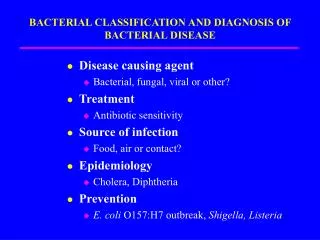
ANTHRAX • Three forms of human anthrax occur: • 1. Cutaneous • 2. Gastrointestinal • Oropharyngeal • Abdominal • 3. Inhalation (Woolsorter’s Disease)
Cutaneous anthrax Vesicle development, day 2 Eschar formation, day 4
Inhalation Anthrax • Infective dose = 8,000 - 15,000 spores • Incubation period = 1-6 days • Duration of illness = 3-5 days • Fever, malaise, and fatigue • Short period of improvement = up to 2 days • Abrupt respiratory distress…death <24hrs • Person to person transmission = no
Anthrax: SpecimenSelection • Inhalation: Sputum and Blood • Cutaneous: Vesicles and Eschar • Gastrointestinal: Stool and Blood
Bacillus anthracisKey Sentinel Lab Tests • Gram stain • Growth characteristics on agar • Sporulation, in air • Motility • Capsule by India Ink
Bacillus anthracisGram Stain Morphology • Broad gram-positive rod: 1-1.5 X 3-5 µ • Oval, central - subterminal spores: 1 X 1.5 µ with no significant swelling of cell • Spores are NOT usually present in clinical specimens unless exposed to atmospheric O2
B. anthracisColonial Morphology • Colonial morphology of 18-24hr @ 35 C: • Well isolated colonies are 2-5 mm in diameter • Flat or slightly convex, irregularly round • Edges: slightly undulate, often curly tailing edges • Ground glass appearance • “Sticky” consistency….stands up like beaten egg whites
Bacillus anthracisPresumptive Identification • Gram-positive, broad rod, catalase-positive, spore-positive, aerobe: Bacillus sp. • Spores are oval and nonswelling with ground glass colony appearance: Bacillus morphology group 1, includes B. anthracis, B. cereus, B cereus var mycoides, and B. thuringiensis
Bacillus anthracisPresumptive Identification, con’t • Nonmotile: B anthracis and B cereus var mycoides (and B. megaterium) • Nonhemolytic, forms capsule: Presumptive B. anthracis • Refer to state lab for testing
Yersinia pestis Plague
Plague: Overview • Natural vector - Rodent flea • Mammalian hosts • rats, squirrels, chipmunks, rabbits, and carnivores • Enzootic or Epizootic
Plague Epidemiology • U.S. averages 13 cases/yr • 30% of cases are in Native Americans in the Southwest. 15% case fatality rate • Most cases occur in summer and near the patient’s residence • bubonic (infected lymph nodes) • septicemic (blood-borne organisms) • pneumonic (transmissible by aerosol; deadliest)
Yersinia pestisSpecimen Selection • Specimen selection is important • Bubo - lymph node aspirate • Blood - organisms may be intermittent. Take three specimens 10-30 minutes apart • Pneumonic • Sputum/throat - use Wayson stain • Bronchial washings - Wayson stain • Inoculate routine plating media
Sentinel Lab ProceduresYersinia pestis • Gram stain • Wayson stain • Growth characteristics on agar • Growth characteristics in broth
Yersinia pestisGram stain • Small, gram-negative coccobacilli
Yersinia pestisWayson Stain • Used for rapid assessment • when it is a part of the identification process • Best with tissue, sputum, blood • Stains of pure culture isolates tend to lose bipolarity • Pink-blue cells with polar granules (safety pin appearance)
Yersinia pestisWayson Stain • Pink-blue cells with a closed safety pin look Wayson stain alone is not diagnostic
Y.pestis 48 h culture on SBA 72 h culture on SBA
Yersinia pestisTechnical Hints • Small Gram-negative, poorly staining rods from blood, lymph node aspirate, or respiratory specimens • Safety pin appearance in Gram, Wright, Giemsa, or Wayson stain • More than one patient in a short, specified period with fever, lymphadenopathy • Refer to state lab
Francisella tularensis Tularemia
Tularemia: Overview • Disease of Northern Hemisphere • In U.S., most cases associated with rabbits/hares and ticks • About 200 cases/year in U.S. • most in South central and Western states • majority of cases in summer, some in winter
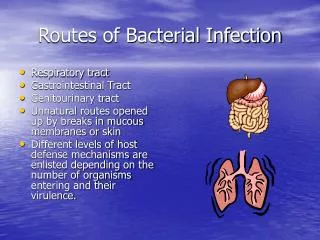
Tularemia: Overview (cont’d) • Several forms of human tularemia exist: • - Ulceroglandular, glandular, oculoglandular, • oropharyngeal, intestinal, pneumonic, and • typhoidal • Low infectious dose • 1 to 10 organisms by aerosol or intradermal route • No person-to-person transmission
Tularemia: Specimen Selection • Serum - acute and convalescent • Blood cultures • Sputum • Swab – ulcer or eye
Sentinel Lab ProceduresFrancisella tularensis • This is a dangerous, highly virulent organism and it should not be manipulated at the bench. Laboratory-acquired infections can occur easily. • Gram stain • Growth characteristics in broth • Growth characteristics in agar
Francisella tularensis • Poorly staining, tiny Gram-negative coccobacilli
Francisella tularensisGrowth Characteristics • Fastidious, requires cysteine for robust growth: Cysteine Heart Agar (CHA) is ideal • Enriched chocolate agar + 9% sheep blood + cysteine • Not part of Sentinel Lab routine procedures • BCYE (for Legionella) also works • Will grow initially on sheep and chocolate blood agar and Thayer-Martin agar, but poorly or not at all on passage • Grows slowly at 35oC, poorly at 28oC
Francisella tularensisGrowth Characteristics • 24 hours • gray-white, translucent colonies • usually too small to be seen individually • 48 hours • Sheep Blood Agar - <1 mm, gray-white, opaque, no hemolysis
Francisellatularensis Sheep blood agar Chocolate agar Cysteine heart agar
Francisella tularensisTechnical Hints If you see: • Tiny, Gram-negative coccobacilli from blood, lymph node aspirate, or respiratory specimens • Blood isolates that will grow slowly on chocolate agar but poorly or not at all on blood agar in 24 hours • Faint growth in thio; requires cysteine in other broth • Refer to state lab
Brucella spp. Brucellosis
BRUCELLOSIS • A zoonotic disease caused by any of 4 Brucella sp.: abortus, melitensis, suis, and canis • A systemic infection characterized by an undulant fever pattern • But relatively rare in the U.S. withapproximately 100 cases/yr
BRUCELLOSIS:TRANSMISSION • Unpasteurized dairy products • The most common mode of transmission • Direct skin contact • Occupational hazard for farmers, butchers, veterinarians, and laboratory personnel • Aerosols • Highly infectious
BRUCELLOSIS • Infective dose = 10 -100 organisms • Incubation period = 5 days - > 6 months • Duration of illness = weeks to months • Fever, profuse sweating, malaise, headache and muscle/back pain. • Person to person transmission = no • Mortality = <5% • Persistence of organism = very stable
Brucella spp.Specimen Selection • Serum • The diagnosis of brucellosis is frequently achieved by serology. An acute & convalescent phase specimen should be collected (21d apart) • Blood or bone marrow • Sources from which Brucellae are most often isolated • Tissue (spleen, liver) • Brucellae occasionally isolated
Brucella spp.Biosafety Alert • Brucellosis is THE most commonly reported laboratory-associated bacterial infection. • Cases have occurred in clinical laboratory settings by “sniffing” cultures, direct skin contact with cultures, and aerosol generating procedures
Sentinel Lab TestsBrucella spp. • Colonial morphology on SBA • Gram stain morphology • Oxidase positive • Urea hydrolysis positive
Brucella spp.Key Sentinel Lab Tests Colonial morphology on SBA • Fastidious • Visible growth may take 48 - 72 hrs • Small (0.5-1.0mm), convex, glistening • Non-hemolytic and non-pigmented
Brucella spp.Key Sentinel Lab Tests Gram Stain Morphology • Tiny (very) • Faintly staining • Gram-negative coccobacilli • 0.5 - 0.7 x 0.6 - 1.5
Brucella spp.Review of Key Tests • Tiny, faintly staining, gram-negative coccobacilli from blood or bone marrow • Slow growth on Sheep Blood Agar, 2-3 days for colony appearance • Oxidase + • Urease + • Handle plates with care • Refer to state lab