Download
1 / 40
410 likes | 641 Views
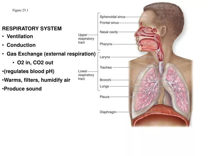
Figure 25.1. RESPIRATORY SYSTEM Ventilation Conduction Gas Exchange (external respiration) O2 in, CO2 out (regulates blood pH) Warms, filters, humidify air Produce sound. Respiratory Tract (and pathway into blood). Nostrils (external nares) Vestibule Nasal cavity Meatuses

E N D
Figure 25.1 RESPIRATORY SYSTEM • Ventilation • Conduction • Gas Exchange (external respiration) • O2 in, CO2 out • (regulates blood pH) • Warms, filters, humidify air • Produce sound
Respiratory Tract (and pathway into blood) Nostrils (external nares) Vestibule Nasal cavity Meatuses Choanae (internal nares) Nasopharynx Oropharynx Laryngopharynx Larynx Rima glottidis/glottis Trachea Primary (main) bronchi Secondary (lobar) bronchi Tertiary (segmental) bronchi Smaller bronchi Bronchioles Terminal bronchioles Respiratory bronchioles Alveolar ducts Alveolar sacs/alveoli blood (in pulmonary capillaries) Respiratory portion Upper tract Lower tract Respiratory membrane
Respiratory Tract & epithelium Nostrils (external nares) Vestibule– strat squam Nasal cavity —resp mucosa Meatuses Choanae (internal nares Nasopharynx—resp mucosa Oropharynx—strat squam Laryngopharynx—strat squam Larynx – strat squam & resp mucosa Rima glottidis Trachea —resp mucosa Primary (main) bronchi —resp mucosa Secondary (lobar) bronchi bronchi —resp mucosa Tertiary (segmental) bronchi bronchi —resp mucosa simple columnar Bronchioles – Terminal bronchioles Respiratory bronchioles – simple cuboidal Alveolar ducts – simple squamous Alveolar sacs/alveoli –simple squam blood simple columnar ↓ simple cuboidal Respiratory membrane – simple squamous x2
PSCC Lamina propria
Figure 25.3 location of paranasal sinuses
Transnasal endoscopy:video of upper respiratory tract http://www.youtube.com/watch?v=wjRsa77u6OU
Gas Exchange is Efficient Due to: Large differences in partial pressures (concentrations) large gradients = fast diffusion Altitude reduces gradientless oxgyenation Small distances short distance across alveolar and capillary wall = fast diffusion edema and pulmonary fibrosis increase distance less oxygenation O2 and CO2 are lipid soluble Overall area of diffusion is great/large (alveolar surface area equal to a tenis court) more surface area = fast diffusion emphasema reduces surface area less oxygenation Pulmonary blood flow is coordinated with ventilation blood is sent to alveoli with most O2 (pulmonary arterioles vasoconstrict in response to low alveolar O2, dilate in response to high alveolar O2)
Coordination of pulmonary blood flow and ventilation: perfusion is matched with ventilation • Pulmonary arterioles dilate in response to high alveolar O2 and constrict in response to low alveolar O2 levels • Results in pulmonary blood flowing to alveoli with high O2 content • Bronchioles dilate in response to high air CO2 levels, constrict in response to low CO2 levels • Results in inhaled air flowing to alveoli that are poorly ventilated
During Inspiration: chest wall expands, diaphragm drops/lowers thoracic cavity vol. increases: pressure decreases Air moves in During Expiration: chest wall retracts, diaphragm moves up thoracic vol. decreases: pressure increases air moves out
During quiet breathing (ventilation) the pressure in the lungs is 1-3 mmHg lower then atmospheric during inspiration During experition it is 1-3 mmHg greater then atmospheric By always being lower then both intrapulmonary and atmsopheric pressure the negative pressure in the pleural cavity (Intrapleural pressure) results in the lungs staying expanded and expanding with the chest wall during inspiration.
During Forced Breathing (ventilation): Accessory muscles are recruited. Intrapulmonary pressure (in lungs/alvoeli) can drop 30 mm Hg below atmospheric and rise 100 mmHg above atmospheric pectoralis minor (not shown)
Fig. 16.13 pectoralis minor (not shown)
Nervous system control Respiratory Centers in brain Medullary Centers: Dorsal Rhymicity Group Stimulates diaphragm during quiet respiration Ventral Rhymicity Group Stimulates accesory muscles needed for forced inhalation and exahalation Pontine Centers Pneumotaxic and apneustic areas Regulate and direct the medulary centers
Sensory Input • Peripheral receptors • Detect O2 • Detect pH (which reflects CO2) • Chemoreceptors in carotid body glossopharyngeal nerve • Chemoreceptors in aortic body vagus nerve • Central Receptors • Detect pH of CSF (a direct indicator of arterial CO2) • Chemosensative Area of Medulla
Table 16.6 This means that the peripheral receptors can be stimulated by pH changes due to factors other than blood PCO2
Fig. 16.26 Nervous System regulation of breathing
Nervous system and breathing • The pons and medulla of brainstem are involved with the regulation of breathing/ventilation: • In the medulla: • The dorsal rhythmicity group is involved with signaling the muscles of inspiration to contract during normal, quiet, resting breathing and during heavily/forced inhalation • Phrenic nerve to diaphragm • Intercostal nerves to external intercostals • The ventral rhymthmicity group signals accessory muscle needed during forced expiration • In The Pons: • The apnuestic and pneumotaxic areas of pons can modify the activity of the DRG and VRG. • You don’t need to know any more details about these two.