Download
1 / 11
130 likes | 275 Views
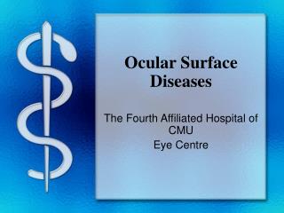
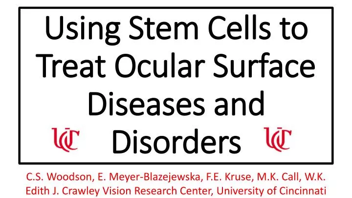
Using Stem Cells to Treat Ocular Surface Diseases and Disorders. C.S. Woodson, E. Meyer- Blazejewska , F.E. Kruse, M.K. Call, W.K. Edith J. Crawley Vision Research Center, University of Cincinnati. Background.

E N D
Using Stem Cells to Treat Ocular Surface Diseases and Disorders C.S. Woodson, E. Meyer-Blazejewska, F.E. Kruse, M.K. Call, W.K. Edith J. Crawley Vision Research Center, University of Cincinnati
Background Limbal epithelial stem cells are located in the limbus, a thin band along the edge of the iris. These cells are responsible for replenishing the cells of the transparent, avascular cornea and separating them from the opaque, conjunctival tissue of the eye that contains blood vessels. Corneal injury or genetic defects can prevent proper cell barrier and replenishment function or destroy them all together, a disorder called Limbal Stem Cell Deficiency (LSCD). As a result, the conjunctiva grows uncontrollably over the clear cornea and causes blindness.
Currently, patients with unilateral LSCD are able to transplant healthy limbal stem cells from the healthy eye to the diseased one to treat LSCD. However, patients with bilateral LSCD cannot do this process and if they accept a transplant from a donor, rejection of the donated cells is possible. • To explore the therapeutic potential of bulge-derived hair follicle stem cells to treat LSCD. Purpose
Hypothesis In this study, we addressed the hypothesis that when transplanted, Hair Follicle Stem Cells (HFSCs) will develop into the corneal epithelial cells needed to treat LSCD. Method • Tri-transgenic mice (K12rtTA /tetO-Cre/RosamTmG) were utilized to harvest HFSCs. Wild-type mice were then given LSCD by the complete removal of all epithelium with a corneal rust-ring remover. The isolated HFSCs were transplanted on a fibrin carrier onto wild-type LSCD mice. Factors in the corneal environment signaled HFSCs to differentiate into corneal cells, exhibited by expression of K12 (in green).
I. Hair Follicle Stem Cell (HFSC) Isolation • Murine HFSCs were isolated, in order to access the bulge region harboring the epithelial stem cells, which express keratin 15 (Krt15) in green.
Results I. HFSC transplant The HFCS from K12rtTA /tetO-Cre/RosamTmGmouse were placed on a wild-type C57/Black6 mouse with induced LSCD (A) and sutured on (B). The eye was then examined post-transplantation under red light (C) and green light (D).
II. Keratin12 (K12) production post-transplant Keratin 12, considered a molecular marker of corneal epithelial cells, is detected with EGFP within 3 days (A) and 2 weeks (B) post-transplantation.
III. HFSC in action HFSCs replaced nonfunctional limbal stem cells and began barrier function and replenishment of avascular cornea once more. A-H are 4 weeks after epithelial tissue was removed (induced LSCD). A-D are images from an LSCD mouse that received a transplants. Images E-H are a control LSCD mouse.
Conclusion • HFSCs can differentiate into corneal epithelial cells. • HFSCs have therapeutic potential for LSCD patients with an 80% success rate. Next Steps Now, we have begun examining environmental factors that cue maintenance and differentiation of limbal stem cells. PPARɣ, a transcription factor, is thought to be one of these factors. This is evident in the human limbus and cornea where PPARɣ density is high in limbus and decreases as you move towards central cornea (A). In mice, PPARɣ is expressed in limbus (B) and only weakly in the central cornea.
(A) (B) Mouse limbus Human limbus Human cornealepithelium Mouse corneal epithelium