Download

1 / 97
990 likes | 1.7k Views
Introduction. Tuberculosis, one of the oldest diseases known to affect humans, is a major cause of death worldwide. This disease, which is caused by bacteria of the Mycobacterium tuberculosis complex? affects the lungs Other organs are involved in up to one-third of cases. . If untreated, the dise

E N D
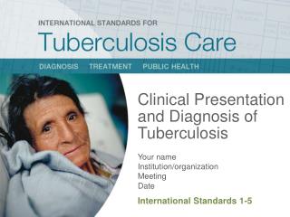
1. Clinical Presentation of Mycobacterium Tuberculosis By Dr.Sujith S.
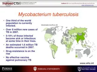
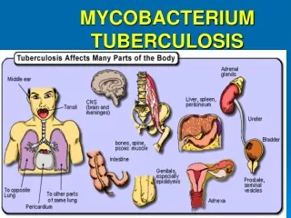
2. Introduction Tuberculosis, one of the oldest diseases known to affect humans, is a major cause of death worldwide.
This disease, which is caused by bacteria of the Mycobacterium tuberculosis complex? affects the lungs
Other organs are involved in up to one-third of cases
3. If untreated, the disease may be fatal within 5 years in 50�65% of cases.
Transmission usually takes place through the airborne spread of droplet nuclei produced by patients with infectious pulmonary tuberculosis.
4. M. bovis (the bovine tubercle bacillus)
M. caprae (related to M. bovis)
M. africanum (isolated from cases in West, Central, and East Africa)
M. microti (the "vole" bacillus, a less virulent and rarely encountered organism)
M. pinnipedii (a bacillus infecting seals and sea lions in the southern hemisphere and recently isolated from humans)
M. canettii
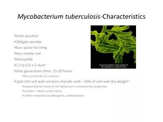
5. Etiological Agent M. tuberculosis is a rod-shaped, non-spore-forming, thin aerobic bacterium measuring 0.5 um by 3 um the bacilli cannot be decolorized by acid alcohol; this characteristic justifies their classification as acid-fast bacilli.
Acid fastness is due mainly to the organisms' high content of mycolic acids, long-chain cross-linked fatty acids, and other cell-wall lipids
6. History Fever
Fatigue
Cough
Weight loss
Dyspnoea
Chest Pain
Retrosternal & interscapular dull pain,worsened by swallowing
7. Cont.d Cough -initially mild,non-productive / productive of only scanty sputum.
May be present only in the morning, when accumulated secretions are expectorated.
Progressive,?cough becomes more continuous and productive of yellow or yellow-green sputum, rarely foul-smelling.
Frank hemoptysis, due to caseous sloughing or endobronchial erosion?present later in the disease.
8. Dyspnoea-occurs when
extensive parenchymal involvement/ pleural effusions, /pneumothorax.
Pleuritic chest pain -when present, signifies inflammation abutting or invading the pleura, with or without an effusion.
Rarely progresses to frank empyema
Patients may present with painful ulcers of the mouth, tongue, larynx or GI tract .
9. Elderly Fever, sweats and hemoptysis were less common in the elderly.
These patients were less likely to have cavitary disease / positive purified protein derivative (PPD) skin test.
10. Cont.d Painless swelling of the lymph nodes, most commonly at posterior cervical and supraclavicular sites.
Increase in urinary frequency
Dysuria
Nocturia
Hematuria
Flank or abdominal pain
11. Pain & swelling in the weight-bearing joints
(the spine in 40% of cases, the hips in 13%, and the knees in 10%).
12. Initially as headache and slight mental changes. May evolve acutely with severe headache, confusion, lethargy, altered sensorium, and neck rigidity.
Paresis of cranial nerves
Involvement of cerebral arteries may produce focal ischemia.
Coma, with hydrocephalus and intracranial hypertension
13. Abdominal pain and swelling
Obstruction
Hematochezia
Palpable mass in the abdomen
14. Diffuse abdominal pain/ pain localizing to the right upper quadrant, nausea, vomiting and diarrhea. Hepatomegaly .
Liver function test abnormalities are common, including elevated alkaline phosphatase and transaminases.
Cholestatic jaundice is also well-documented in miliary TB.
Ascites may be detected in occasional patients. Miliary TB may also present as pancreatitis / cholecystitis .
15. Physical Examination Not specific and usually are absent in mild or moderate disease.
Dullness with decreased fremitus & resonance may indicate pleural thickening or effusion.
Rales may be present throughout inspiration, or may be heard only after a short cough (post-tussive rales).
16. When large areas of the lung are involved, signs of consolidation associated with open bronchi? whispered pectoriloquy/tubular breath sounds.
Distant hollow breath sounds over cavities are called amphoric,after the sound made by blowing across the mouth of jars used in antiquity (amphora).
17.
Pulmonary Tuberculosis
18. Primary Disease Primary pulmonary tuberculosis occurs soon after the initial infection with tubercle bacilli.
Inspired air is distributed to the middle and lower lung zones-most commonly involved in primary tuberculosis.
The lesion -accompanied in more than half of cases by hilar or paratracheal lymphadenopathy.
The lesion heals spontaneously ,later be evident as a small calcified nodule (Ghon lesion).
19. Post Primary disease Chronic TB
Postprimary disease
Recrudescent TB
Endogenous reinfection
Adult type progressive TB
20. Reactivation of a previously dormant focus seeded at the time of the primary infection.
The apical posterior segments of the lung are frequently involved.
The original site of spread may have been previously visible as a small scar called a Simon focus
21. Localized to the apical and posterior segments of the upper lobes- higher mean oxygen tension (compared with that in the lower zones) favors mycobacterial growth.
The superior segments of the lower lobes are frequently involved.
Small infiltrates to extensive cavitary disease..
22. Liquefied necrotic contents - discharged into the airways? satellite lesions within the lungs that may in turn undergo cavitation
Massive involvement of pulmonary segments or lobes, with coalescence of lesions, produces tuberculous pneumonia.
Some pulmonary lesions become fibrotic and may later calcify, but cavities persist in other parts of the lungs
23. Endobronchial TB
Direct extension to the bronchi from an adjacent parenchymal focus, usually a cavity.
Spread of organisms to the bronchi via infected sputum from a distant site.
24. Clinical Presentation
Bronchorrhea, which is production of more than 500 mL per day of sputum .
Caseous material from endobronchial lesions / calcific material from extension of calcific nodes into the bronchi can be expectorated, which is known as lithoptysis.
25. Wheezing and hemoptysis may also be seen.
Lymph node rupture can be associated with chest pain.
Dyspnea may signal obstruction or atelectasis
Physical findings
Diminished breath sounds, rhonchi or wheezing may be heard.
The wheeze is described as low-pitched, constant and always heard over the same area on the chest wall.
26. �Lower lung field TB
Disease located below a line traced across the hila, including the perihilar regions, on a standard PA and lateral chest x-ray.
Risk factors:
Elderly patients
Diabetes
Renal/ hepatic disease
Those receiving corticosteroids
Underlying silicosis appear most at risk for lower lobe
28. Complications of Pulmonary TB
Hemoptysis
Pneumothorax
Bronchiectasis
Extensive pulmonary destruction
29. Hemoptysis
Active disease(smear positive)
"Rasmussen's aneurysm" causes massive hemoptysis when TB extends into the adventitia and media of bronchial arteries, resulting in inflammation and thinning of the vessel wall.
Subsequently rupturing into the cavity, producing hemoptysis.
30. Other causes
residual bronchiectasis
an aspergilloma/ other fungus ball invading an old healed cavity,
a ruptured broncholith that erodes through a bronchial artery
a carcinoma
another infectious or inflammatory process.
31.
CNS Tuberculosis
32. Meningitis
Intracranial tuberculoma
Spinal tuberculous arachnoiditis
33. Pathogenesis Scattered tuberculous foci (tubercles) are established in the brain, meninges, or adjacent bone during the bacillemia.
The chance occurrence of a subependymal tubercle, with progression and rupture into the subarachnoid space.
34. The spillage of tubercular protein into the subarachnoid space produces an intense hypersensitivity reaction.
35. Proliferative arachnoiditis, most marked at the base of the brain, produces a fibrous mass involving cranial nerves and penetrating vessels.
Communicating hydrocephalus results from extension of the inflammatory process to the basilar cisterns and impedance of CSF circulation and resorption.
36. Vasculitis with resultant thrombosis and infarction involves vessels that traverse the basilar/ spinal/ are located within the brain substance.
37. Meningitis
?The prodromal phase(2 to 3 weeks)
fever,malaise,lassitude
?The meningitic phase-
meningismus, protracted headache, vomiting, lethargy, confusion, and varying degrees of cranial nerve and long-tract signs
38. The paralytic phase:
?delirium, stupor, coma, seizures, multiple cranial nerve palsies,or dense hemiplegia.
39. About one-third of patients on presentation ?generalized (miliary) tuberculosis
Careful fundoscopic examination often shows choroidal tubercles .
These are multiple, ill-defined, raised yellow-white nodules (granulomas) of varying size near the optic disk.
40. Tuberculoma
Tuberculomas are conglomerate caseous foci within the substance of the brain ? from deep-seated tubercles acquired during a recent or remote hematogenous bacillemia.
41. Radiologically
clinically silent
single / multiple enhancing nodular lesions , on CT scanning of patients with meningitis or miliary TB without meningitis.
42. Tuberculoma is also a distinct clinical syndrome manifested by focal neurologic symptoms and signs of an intracranial mass lesion.
43.
Tuberculosis Lymphadenitis
44. Pathogenesis Isolated peripheral tuberculous lymphadenopathy ?reactivation of disease at a site seeded hematogenously during primary TB infection.
45. Clinical Presentation The most common presentation is isolated chronic, nontender, lymphadenopathy. The mass may be present for up to 12 months before diagnosis
A firm discrete mass or matted nodes fixed to surrounding structures; the overlying skin may be indurated. Uncommon findings include fluctuance, draining sinus/ erythema nodosum
46. Cervical lymphadenopathy is the most common manifestation of TB lymphadenitis.
A unilateral mass appears in the anterior or posterior cervical triangles; submandibular and supraclavicular lymph node involvement also occurs.
Bilateral disease is uncommon.
Multiple nodes may be involved at that site.
47. The axillary, inguinal, mesenteric, mediastinal, and intramammary lymph nodes.
Mediastinal lymphadenopathy
Dysphagia
Esophageal perforation
Vocal cord paralysis due to recurrent laryngeal nerve involvement
Pulmonary artery occlusion mimicking pulmonary embolism.
48. Tuberculous peritoneal lymphadenopathy ?i)periportal region
?ii)peripancreatic
?iii)mesenteric lymph nodes.
?iv)Hepatic lymph node involvement can lead to jaundice, portal vein thrombosis, and portal hypertension.
Extrinsic compression of renal arteries due to tuberculous abdominal lymphadenopathy can result in renovascular hypertension
49.
Skeletal Tuberculosis
50. Spinal TB (Pott's disease) most often affects the lumbar and lower thoracic region. Tuberculous abscess ? frequently bilateral.
Tuberculous arthritis tends to occur in the weight-bearing joints, the hip and the knee, and is usually monoarticular.
51. Pathogenesis The bacillemia
Seeds organisms
Bone and synovium
52. Spinal TB
Infection - in the anteroinferior aspect of the cancellous vertebral body, inflammatory bone destruction and caseating necrosis.
Active infection spreads down behind the anterior ligament , involves the adjacent vertebral body.
Local destruction - collapse of bony structures, herniation of the disk into the vertebral bodies.
53. A tuberculous abscess - impinge on local structures.
An epidural abscess -produce pressure on the cord.
An extraspinal soft tissue mass - erodes ribs and adjacent structures
Psoas abscess - track down to the groin
54. Vertebral collapse
spinal instability with gibbous deformity
distort the anatomy of the canal to the point of spinal cord compression.
Granulomatous changes ?synovial proliferation in tuberculous arthritis with joint effusion and erosion of cartilage.
Slowly progressive destruction, disorganization of joint architecture, and potential deformity.
55. Clinical Presentation Local pain-associated with muscle spasm and rigidity.
Description of the patient with spinal TB reads: {"The muscle spasm, which extends beyond the diseased area, sometimes produces the well-known erect posture and 'aldermanic' gait}.
The patient walks with short deliberate steps to avoid any jarring of the spine.
56. ii)Cord compression during the active phase of the infection resulting in paraplegia (Pott's paraplegia)
Late-onset paraplegia ?osteophytes and other chronic degenerative changes at a site of previous infection.
TB of the pedicles and other posterior spinal elements is well-described.
57.
Renal Tuberculosis
58. Spinal TB
59. TB Arthritis of Hip
60. Two major mechanism:
i)Direct infection of the kidney and lower urinary tract
ii)Secondary amyloidosis.
iii)Nephrotoxicity induced by antimicrobial agents
iv)Hyponatremia due to the syndrome of inappropriate ADH secretion because of pulmonary involvement /miliary disease.
61. Pathogenesis Hematogenous seeding either at the time of primary pulmonary infection.
The net effect is the formation of granulomatous lesions in the glomeruli ? heal without producing any renal disease.
The granulomas may caseate and rupture into the tubular lumen as late as 30 years after the initial infection.
Tubercle bacilli can enter the medullary interstitium, eventually leading to granuloma formation and progressive medullary injury.
Similar lesions can occur in the ureters, bladder, and, in males, the prostate and epididymis.
62. Dysuria and gross hematuria being the most common symptoms .
Renal colic ? uncommon manifestation with active extrarenal disease (fever, weight loss, cough, hemoptysis).
Rupture of the glomerular granulomas is a random event, independent of disease elsewhere
.
63. Incidentally found to have pyuria and/or microscopic hematuria.
Heavy proteinuria and cellular casts.
Plasma creatinine concentration is usually normal or near normal.
Ureteral strictures may cause obstructive uropathy-> severe cases, renal loss .
64. Signs of extrarenal disease include ureteral strictures (single or multiple)
A contracted bladder
Calcifications in the vas deferens, seminal vesicles, or prostate.
65. Refractory hypertension ? complication of renal tuberculosis.
The elevation in blood pressure ? is angiotensin II-mediated-from intimal proliferation of vessels in or near areas of inflammation, leading to segmental ischemia and renin release.
66. Drug induced Nephritis
Renal glucosuria
Hyperuricosuria
polyuria due to nephrogenic diabetes insipidus
increased urinary excretion of polyclonal light chains.
67. Mild hyponatremia (plasma sodium concentration usually between 125 and 135 meq/L) .
Most patients appear to have the syndrome of inappropriate ADH secretion.
68.
Tuberculosis Peritonitis
69. Risk factors
Cirrhosis
HIV infection
Diabetes mellitus
Underlying malignancy?treatment with anti-tumor necrosis factor (TNF) agents
Patients undergoing continuous ambulatory peritoneal dialysis
70. Reactivation of latent tuberculous foci in the peritoneum ? hematogenous spread from a primary lung focus.
As the disease progresses-? the visceral and parietal peritoneum become increasingly studded with tubercles.
Ascites develops secondary to "exudation" of proteinaceous fluid from the tubercles.
71. Clinical Presentation Ascites (93 percent), abdominal pain (73 percent), and fever (58 percent).
Many patients had a diffusely distended tender abdomen.
The classic doughy abdomen is associated with the fibroadhesive form of tuberculous peritonitis.
72. The salient clinical findings :
Abdominal tenderness
Hepatomegaly
Ascites
73.
Genital Tuberculosis
74. Tuberculosis - affect the upper female reproductive tract (fallopian tube, endometrium, ovaries)
i)Extension from an intraabdominal focus, hematogenous seeding
ii)Ascending from lower genital tract (cervix, vagina, vulva) infection.
The fallopian tube and endometrium are most commonly involved.
75. The finding of endometrial tuberculosis always means that the tubes are infected
Tuberculous salpingitis can exist without associated endometritis.
Pelvic mass, infertility, abnormal uterine bleeding, and pelvic pain
Epididymo-orchitis in males
76.
Tuberculosis Pericarditis
77. Stage of infection
The degree of pericardial involvement
The degree of extrapericardial tuberculous disease
78. Physical Findings
Fever
Tachycardia
Increased jugular venous pressure
Hepatomegaly
Ascites
Peripheral edema
Pericardial rub and distant heart sounds
79. Constructive Pericarditis
Kussmaul's sign (the lack of an inspiratory decline in jugular venous pressure).
Elevated and distended jugular veins with a prominent Y descent (the second inward deflection of the internal jugular pulse due to diastolic inflow of blood into the right ventricle)
In constriction, the inspiratory decline in pressure is not transmitted to the right heart chambers.
80. Cardiac Tamponade
Hypotension with a narrow pulse pressure, reflecting the limited stroke volume.
Sinus tachycardia
Elevated jugular venous pressure
Pulsus paradoxus, defined as an abnormally large decrease in systolic blood pressure (>10 mmHg) on inspiration.
81. Ascites .
The main clue? elevation of jugular venous pressure.
82.
Tuberculosis Enteritis
83. Pathogenesis Swallowing of infected sputum
Hematogenous spread from active pulmonary or miliary TB
Ingestion of contaminated milk or food, a rarity in the Western world
Contiguous spread from adjacent organs
84. Most common site:ileocecal region
Cause:
Relative stasis and abundant lymphoid tissue. The organism penetrates the mucosa and localizes in the submucosal lymphoid tissue, where it initiates an inflammatory reaction with subsequent lymphangitis, endarteritis, granuloma formation, caseation necrosis, mucosal ulceration, and scarring.
85. Ulcerative (60 percent)?multiple superficial ulcers.
Hypertrophic (10 percent)? scarring, fibrosis, and pseudotumor lesions.
Ulcerohypertrophic (30 percent)? inflammatory mass centering around the ileocecal valve with thickened and ulcerated intestinal walls.
86. Clinical Presentation Nonspecific chronic abdominal pain
Anorexia, fatigue, fever, night sweats, weight loss, diarrhea, constipation, or blood in the stool may be present.
A palpable right lower quadrant abdominal mass is present in 25 to 50 percent of patients.
Small bowel obstruction�/ colonic perforation . The presence of ascites may help to distinguish ileocecal TB from Crohn's disease.
87.
Tb Pleural Effusion
88. Pathogenesis Tuberculous pleural effusions ? a delayed hypersensitivity reaction to mycobacterial antigens in the pleural space.
These antigens probably enter the pleural space after rupture of a subpleural focus of disease or leakage from these lesions.
89. Clinical Presentation Tuberculous pleural effusion ? acute febrile illness causing a i) nonproductive cough (94 percent) ii) pleuritic chest pain (78 percent) without an elevation in the peripheral white blood cell (WBC) count.
Night sweats, chills, weakness, dyspnea, and weight loss.
90.
TB Adrenalitis
91. Tuberculous adrenalitis results from hematogenous spread from active infection elsewhere in the body .
Extraadrenal tuberculosis is usually evident, but may be clinically latent
92.
Miliary Tuberculosis
93. Clinical Presentation During primary infection, the disease has a more acute onset and more rapid clinical course.
Acute disease may be fulminant,
i) multiorgan system failure
ii)a syndrome of septic shock
iii) acute respiratory distress syndrome, ARDS
94. Late miliary tuberculosis can be acute, but is more likely to be subacute or chronic.
Reinfection may have a role in highly endemic areas.
At the chronic end of the spectrum, patients may present with failure to thrive without fever, with fever of unknown origin, or with dysfunction of one organ system
96.
Thankyou