Download
1 / 20
200 likes | 366 Views
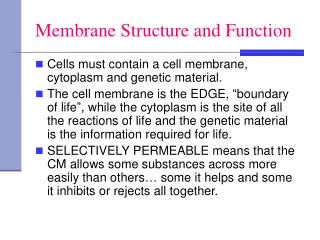
Membrane Structure and Function. © 2011 Pearson Education, Inc. Overview: Life at the Edge. Plasma membrane is boundary that separates living cell from surroundings Plasma membrane is selective permeability. © 2011 Pearson Education, Inc.

E N D
© 2011 Pearson Education, Inc. Overview: Life at the Edge • Plasma membrane is boundary that separates living cell from surroundings • Plasma membrane is selective permeability
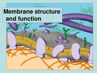
© 2011 Pearson Education, Inc. Cellular membranes are fluid mosaics of lipids and proteins • Phospholipids • most abundant lipid in plasma membrane • amphipathicmolecules • Fuid mosaic model: • membrane is a fluid structure with “mosaic” of various embedded proteins
Figure 7.2 WATER Hydrophilichead Hydrophobictail WATER
Figure 7.3 Phospholipidbilayer Hydrophobic regionsof protein Hydrophilicregions of protein
© 2011 Pearson Education, Inc. • Freeze-fracture studies of the plasma membrane supported the fluid mosaic model • Freeze-fracture is specialized preparation technique that splits membrane along the middle of phospholipid bilayer
Figure 7.4 TECHNIQUE Extracellularlayer Proteins Knife Plasma membrane Cytoplasmic layer RESULTS Inside of cytoplasmic layer Inside of extracellular layer
© 2011 Pearson Education, Inc. The Fluidity of Membranes • Phospholipids membrane can move within bilayer • Most of the lipids, and some proteins, drift laterally • Rarely does a molecule flip-flop transversely across the membrane
Figure 7.5 Fibers of extra-cellular matrix (ECM) Glyco-protein Carbohydrate Glycolipid EXTRACELLULARSIDE OFMEMBRANE Cholesterol Microfilamentsof cytoskeleton Peripheralproteins Integralprotein CYTOPLASMIC SIDEOF MEMBRANE
Figure 7.6 Flip-flopping across the membraneis rare ( once per month). Lateral movement occurs107 times per second.
Figure 7.7 RESULTS Membrane proteins Mixed proteinsafter 1 hour Mouse cell Human cell Hybrid cell
Figure 7.8 Viscous Fluid Unsaturated hydrocarbontails Saturated hydrocarbon tails (a) Unsaturated versus saturated hydrocarbon tails (b) Cholesterol within the animal cell membrane Cholesterol
© 2011 Pearson Education, Inc. Membrane Proteins and Their Functions • Peripheral proteins bound to surface of membrane • Integral proteins penetrate the hydrophobic core • Integral proteins that span the membrane are called transmembrane proteins • The hydrophobic regions of an integral protein consist of one or more stretches of nonpolar amino acids, often coiled into alpha helices
Figure 7.9 EXTRACELLULARSIDE N-terminus helix C-terminus CYTOPLASMICSIDE
© 2011 Pearson Education, Inc. • Six major functions of membrane proteins • Transport • Enzymatic activity • Signal transduction • Cell-cell recognition • Intercellular joining • Attachment to the cytoskeleton and extracellular matrix (ECM)
Figure 7.10 Signaling molecule Receptor Enzymes ATP Signal transduction (a) Transport (c) Signal transduction (b) Enzymatic activity Glyco-protein (f) Attachment to the cytoskeleton and extracellular matrix (ECM) (e) Intercellular joining (d) Cell-cell recognition
Figure 7.10a Signaling molecule Receptor Enzymes ATP Signal transduction (c) Signal transduction (b) Enzymatic activity (a) Transport
Figure 7.10b Glyco-protein (f) Attachment to the cytoskeleton and extracellular matrix (ECM) (e) Intercellular joining (d) Cell-cell recognition
© 2011 Pearson Education, Inc. The Role of Membrane Carbohydrates in Cell-Cell Recognition • Cells recognize each other by binding to surface molecules, often containing carbohydrates, on the extracellular surface of the plasma membrane • Membrane carbohydrates may be covalently bonded to lipids (forming glycolipids) or more commonly to proteins (forming glycoproteins) • Carbohydrates on the external side of the plasma membrane vary among species, individuals, and even cell types in an individual
Figure 7.11 HIV Receptor(CD4) Receptor (CD4)but no CCR5 Plasmamembrane Co-receptor(CCR5) HIV can infect a cell thathas CCR5 on its surface,as in most people. HIV cannot infect a cell lackingCCR5 on its surface, as in resistant individuals.