Download
1 / 86
860 likes | 868 Views
Cell Membranes. 6 Cell Membranes. 6.1 What Is the Structure of a Biological Membrane? 6.2 How Is the Plasma Membrane Involved in Cell Adhesion and Recognition? 6.3 What Are the Passive Processes of Membrane Transport? 6.4 What Are the Active Processes of Membrane Transport?

E N D
6 Cell Membranes • 6.1 What Is the Structure of a Biological Membrane? • 6.2 How Is the Plasma Membrane Involved in Cell Adhesion and Recognition? • 6.3 What Are the Passive Processes of Membrane Transport? • 6.4 What Are the Active Processes of Membrane Transport? • 6.5 How Do Large Molecules Enter and Leave a Cell?
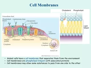
6 Cell Membranes The cell membrane regulates what enters and leaves the cytoplasm. Some cell membranes have pores called aquaporins that allow water to pass freely. Opening Question: Water purity is a worldwide problem. Can aquaporin membrane channels be used in water purification?
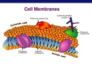
6.1 What Is the Structure of a Biological Membrane? • The general structure of biological membranes is known as the fluid mosaic model. • Phospholipids form a bilayer, which is like a “lake” in which a variety of proteins “float.”
6.1 What Is the Structure of a Biological Membrane? • Phospholipids have a polar, hydrophilic “head” and hydrophobic fatty acid “tails.” • In an aqueous environment, phospholipids form a bilayer.
6.1 What Is the Structure of a Biological Membrane? • Artificial bilayers can be made in the laboratory. • Lipids maintain a bilayer organization spontaneously. This helps membranes fuse during phagocytosis, vesicle formation, etc.
6.1 What Is the Structure of a Biological Membrane? • Lipid composition of membranes vary. • Phospholipids vary in fatty acid chain length, degree of saturation, and phosphate groups.
6.1 What Is the Structure of a Biological Membrane? • Animal cell membranes may be up to 25% cholesterol, which is important for membrane integrity.
6.1 What Is the Structure of a Biological Membrane? • The fatty acid tails make the interior somewhat fluid, allowing lateral movement of molecules. • Fluidity depends on temperature and lipid composition.
6.1 What Is the Structure of a Biological Membrane? • Cholesterol and long-chain, saturated fatty acids pack tightly, making a less-fluid membrane. • As temperature decreases, movement of molecules and cellular processes slow. Some organisms change the lipid content of the cell membranes when they get cold.
6.1 What Is the Structure of a Biological Membrane? • Membranes also contain proteins; the number varies depending on membrane function. • Peripheral membrane proteins lack exposed hydrophobic groups and do not penetrate the bilayer.
6.1 What Is the Structure of a Biological Membrane? • Integral membrane proteins have hydrophobic and hydrophilic regions or domains. • Some extend across the lipid bilayer; others are partially embedded.
6.1 What Is the Structure of a Biological Membrane? • Freeze-fracturing is a technique that reveals proteins embedded in the phospholipid bilayers of cellular membranes.
Figure 6.4 Membrane Proteins Revealed by the Freeze-Fracture Technique
6.1 What Is the Structure of a Biological Membrane? • The proteins and lipids interact noncovalently. • But some membrane proteins have lipid groups covalently attached and are tethered to the lipid bilayer.
6.1 What Is the Structure of a Biological Membrane? • Transmembrane proteins extend all the way through the phospholipid bilayer. • They have one or more transmembrane domains, and the domains on the inner and outer sides of the membrane can have specific functions. • Peripheral membrane proteins are located on one side of the membrane.
6.1 What Is the Structure of a Biological Membrane? • Some membrane proteins can move freely within the bilayer, while some are anchored to a specific region. • When cells are fused experimentally, some proteins from each cell distribute themselves uniformly around the membrane.
6.1 What Is the Structure of a Biological Membrane? • Membranes are dynamic and are constantly forming, transforming, fusing, and breaking down.
6.1 What Is the Structure of a Biological Membrane? • Membranes also have carbohydrates on the outer surface that serve as recognition sites for other cells and molecules. • Glycolipids—carbohydrate + lipid • Glycoproteins—carbohydrate + protein
Working with Data • A key experiment providing evidence for the fluid mosaic model used the technique of cell fusion to show that membrane proteins rapidly diffuse within the cell membrane.
Working with Data 6.1: Rapid Diffusion of Membrane Proteins • Question 1: • Plot the percentage of fully mixed cells over time. • How long did it take for complete mixing?
Working with Data 6.1: Rapid Diffusion of Membrane Proteins • Question 2: • What does your answer to Question 1 indicate about the rate of diffusion of the mouse and human proteins?
6.2 How Is the Plasma Membrane Involved In Cell Adhesion and Recognition? • Cells arrange themselves in groups by cell recognition and cell adhesion. • These processes can be studied in sponge cells—the cells are easily separated and will come back together again.
6.2 How Is the Plasma Membrane Involved In Cell Adhesion and Recognition? • Molecules involved in cell recognition and binding are glycoproteins. • Binding of cells is usually homotypic: The same molecule sticks out from both cells and forms a bond. • Some binding is heterotypic: The cells have different proteins.
6.2 How Is the Plasma Membrane Involved In Cell Adhesion and Recognition? • Cell junctions are specialized structures that hold cells together: • • Tight junctions • • Desmosomes • • Gap junctions
Figure 6.7 Junctions Link Animal Cells Together (Part 1) • Tight junctions help ensure directional movement of materials.
Figure 6.7 Junctions Link Animal Cells Together (Part 2) • Desmosomes are like “spot welds.”
Figure 6.7 Junctions Link Animal Cells Together (Part 3) • Gap junctions allow communication.
6.2 How Is the Plasma Membrane Involved In Cell Adhesion and Recognition? • Cell membranes also adhere to the extracellular matrix. • The transmembrane protein integrin binds to the matrix outside epithelial cells, and to actin filaments inside the cells. • The binding is noncovalent and reversible.
6.2 How Is the Plasma Membrane Involved In Cell Adhesion and Recognition? • Cells can move within a tissue by the binding and reattaching of integrin to the extracellular matrix. • This is important for cell movement within developing embryos and for the spread of cancer cells.
6.3 What Are the Passive Processes of Membrane Transport? • Membranes have selective permeability—some substances can pass through, but not others. • Passivetransport—no outside energy required (diffusion). • Activetransport—energy required.
6.3 What Are the Passive Processes of Membrane Transport? • Energy for passive transport comes from the concentration gradient: the difference in concentration between one side of the membrane and the other.
6.3 What Are the Passive Processes of Membrane Transport? • Particles in a solution move randomly until they are evenly distributed. • At equilibrium, the particles continue to move, but there is no net change in distribution.
6.3 What Are the Passive Processes of Membrane Transport? • Diffusion: the process of random movement toward equilibrium. • Net movement is directional until equilibrium is reached. • Diffusion is the net movement from regions of greater concentration to regions of lesser concentration.
6.3 What Are the Passive Processes of Membrane Transport? • Diffusion rate depends on: • Diameter of the molecules or ions • Temperature of the solution • Concentration gradient
6.3 What Are the Passive Processes of Membrane Transport? • Diffusion works very well over short distances (e.g., within a cell). • Membrane properties affect the diffusion of solutes. • A membrane is permeable to solutes that move easily across it; impermeable to those that cannot.
6.3 What Are the Passive Processes of Membrane Transport? • Simple diffusion: Small molecules pass through the lipid bilayer. • Water and lipid-soluble molecules can diffuse across the membrane. • Electrically charged and polar molecules can not pass through easily.
6.3 What Are the Passive Processes of Membrane Transport? • Osmosis: the diffusion of water. • It depends on the relative concentrations of water molecules on each side of the membrane. • Hypertonic: higher solute concentration • Isotonic: equal solute concentrations • Hypotonic: lower solute concentration