Download
1 / 37
370 likes | 379 Views
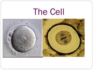
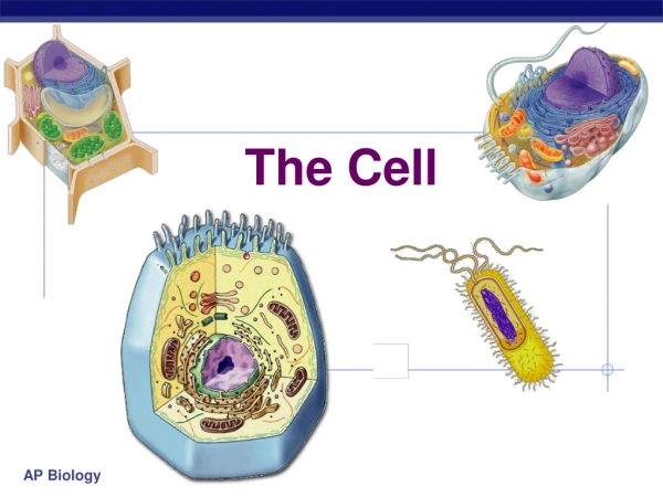
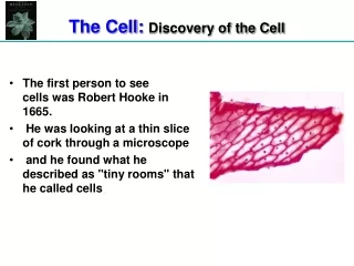
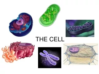
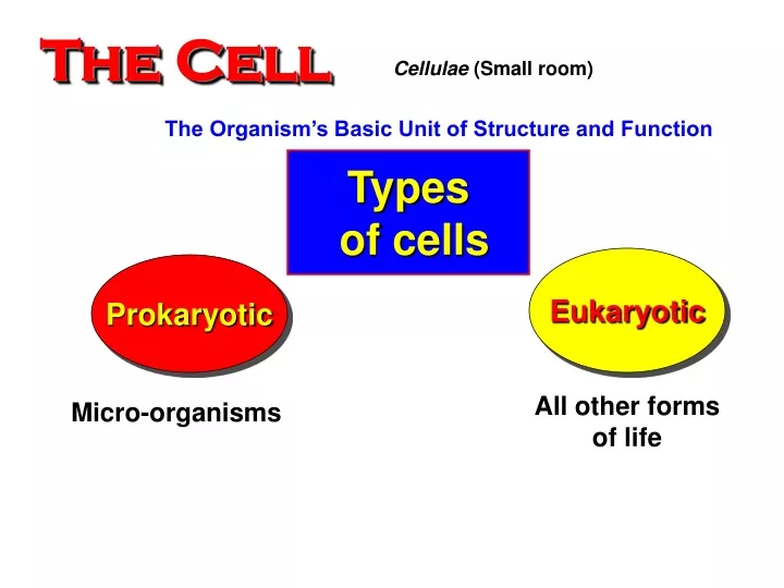
The Cell. Cellulae (Small room). The Organism’s Basic Unit of Structure and Function. Types of cells. Eukaryotic. Prokaryotic. All other forms of life. Micro-organisms. Cell Theory. 1- All organisms are composed of one or more of cells. 2- Cell is the basic unit of life.

E N D
The Cell Cellulae (Small room) The Organism’s Basic Unit of Structure and Function Types of cells Eukaryotic Prokaryotic All other forms of life Micro-organisms
Cell Theory 1- All organisms are composed of one or more of cells. 2- Cell is the basic unit of life. 3- The new cell arises only from pre- existing cell.
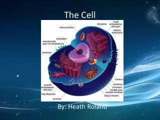
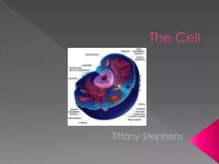
All cells are surrounded by a plasma membrane. The semi-fluid substance within the cell is called “cytosol”, containing the cell organelles. All cells contain chromosomes which have genes in the form of DNA. All cells have tiny organelles called “Ribosomes” that make proteins. 1). Prokaryotic and eukaryotic cells differ in size and complexity Similarities
A major difference between prokaryotic and eukaryotic cells is the location of chromosomes. In an eukaryotic cell, chromosomes are contained in a true nucleus ). In a prokaryotic cell, the DNA is concentrated in the nucleoid) without a membrane separating it from the rest of the cell. In prokaryotic cell, DNA is a single strand or double strand DNA. But in eukaryotic cell, DNA is double strand. 1). Prokaryotic and eukaryotic cells differ in size and complexity Differences
What are Prokaryotes? • It includes two Major Domains: Archaea and Bacteria • Prokaryotes are single-celled organisms that do not have a membrane-bound nucleus, and can live in nearly every environment on Earth. • Although tiny, prokaryotes differ greatly in their genetic traits, their modes of nutrition, however, their habitats are similar. • Based on genetic differences, prokaryotes are grouped in two domains: Domain Archaea and Domain Bacteria.
Prokaryotes • “Prokaryote” means “before a nucleus” • No internal membrane-bound organelles – just one little bag of cytoplasm • No nucleus • Usually single-celled (may form simple colonies) • May or may not require oxygen for survival. • Earliest types of cells on Earth • Cell type of all bacteria and Archaea
Much tougher than eukaryotes Can survive almost anywhere – and do! Have much greater genetic diversity than eukaryotes Have a cell wall surrounding the cell membrane (different chemistry from plant cell wall)
Types of Prokaryotes Prokaryotes Bacteria Archaea - Exist in extreme environments (hot and salty) - Exist in most environments They are differing in some other structural, biochemical and physiological characteristics
1. Domain: Archaea Archaea are extremophiles, “مُحب للظروف القاسية” of extreme environments and can be classified into: a)- Extreme halophilesمُحب للملوحة: live in such saline places as the Great Salt Lake and the Dead Sea. Some species require an extremely saltyشديدة الملوحة environment to grow. b)- Extreme thermophilesمُحب للحرارة live in hot environments. The optimum temperatures for most thermophiles are 60 - 80°C.
شبه نواة الريبوزومات غشاء بلازمى الجدار الخلوى الكبسولة الأسواط 2. Domain: Bacteria Bacteria occur in many shapes and sizes. Bacteria of four shapes: rod-shaped, sphere-shaped, spiral-shaped, or filamentous-shaped. الأهداب
Plasma membrane Cell Wall Capsule Ribosomes Nucleoid Cytoplasm (Cytosol) Prokaryotic Cell
Shapes of Bacteria • Bacteria occur in many shapes and sizes. Most bacteria have one of three basic shapes: rod-shaped, sphere-shaped, or spiral-shaped. • Spiral shaped bacteria in the form of spirilla (singular, spirillum) or vibrio (comma like). • Sphere-shaped bacteria are called cocci (singular, coccus). An example of cocci is Micrococcus luteus. Cocci are single or aggregate cells in different shapes. • Rod-shaped bacteria are called bacilli (singular, bacillus). An example of bacilli is Escherichia coli. Bacilli are single or aggregate cells in different shapes also.
It is a tool for identifying تعريف bacteria, based on differences in their cell walls. A)- Gram-positive (Gram +ve) bacteria: Their cell walls have large amountsكمية كبيرةof peptidoglycans that react with Gram’s stain (appear violet-stained تـُصبغ بنفسجيا). The Gram’s stain: صبغة جرام
B)- Gram-negative(Gram -ve) bacteria: their cell walls have no or small amount of peptidoglycan. So, do not react or very weakly react withGram’s stain (appear red-stained تصبغ بالأحمر) The Gram’s stain: صبغة جرام
Summary of Gram’s stain: صبغة جرام • Gram Stain • Most species of bacteria are classified into two categories based on the structure of their cell walls as determined by a technique called the Gram stain. • Gram-positive bacteriahave a thick layer of peptidoglycan in their cell wall, and they appear violet under a microscope after the Gram-staining procedure. • Gram-negative bacteriahave a thin layer of peptidoglycan in their cell wall, and they appear reddish-pink under a microscope after the Gram-staining procedure.
Gram +ve bacteria: have Large amount of peptidoglycan that stained violet. Gram –ve bacteria: Have small amount or no peptidoglycan stainedred. Summary of Gram’s stain: صبغة جرام • Most Gram-negative species are pathogenic (ممرضة ) more threatening (أكثر خطورة) than gram-positive species. • Gram-negative bacteria are commonly more resistant (أكثر ممانعة) than gram-positive species to antibiotics للمضادات الحيوية.
I - the bacterial capsule • Many prokaryotes (bacteria) secrete a sticky protective layer called capsule outside the cell wall. • Capsule has the following functions وظائف: • Adhere تثبيت bacterial cells to their substratum السطح. • Increase bacterial resistance المقاومة to host defenses مناعةالعائل. • Stickتلصق)) bacterialcells togetherwhen live incolonies. • Protect تحمى bacterial cell.
II - The bacterial cell wall • In all prokaryotes, the functions of the cell wall are as following: • maintains تحافظ the shape of the cell, • affords physical protection الحماية الطبيعيةتوفر • prevents the cell from bursting (إنفجار) in a hypotonic environment البيئة ذات التركيز الأسموزى المنخفض. • Most bacterial cell walls contain peptidoglycan (a polymer of modified sugars cross-linked by short polypeptides). • The walls of Archaea lack (تـفـتـقـد) peptidoglycan.
Nutritional and metabolic diversity All prokaryotes (and eukaryotes too) are grouped into four (4) categories according to how they obtain energy and carbon. 1. Photoautotrophs - Photosynthetic à use light as the energy source - CO2 is the carbon source Example: Cyanobacteria; plants (eukaryotic).
2. Chemoautotrophs - Energy from oxidation of inorganic substances (e.g. NH4, and S) - CO2 is the carbon source Example: Sulfolobus, Beggiatoa (shown on slide)
3. Photoheterotrophs - Light as energy source - Organic compounds are source of carbon 4. Chemoheterotrophs - Organic compounds are energy source and source of carbon (this includes humans) Examples: Many prokaryotes; animals (eukaryotic); fungi (eukaryotic)
binary fission in bacteria • Cell division involves inward growth of the plasma membrane, dividing the parent cell into two daughter cells, each with a complete genome.
In eukaryote cells, the chromosomes are contained within a membranous nuclear envelope. • The region between the nucleus and the plasma membrane is the cytoplasm. • All the material within the plasma membrane of a prokaryotic cell is cytoplasm. • Within the cytoplasm of a eukaryotic cell is a variety of membrane-bounded organelles of specialized form and function. • These membrane-bounded organelles are absent in prokaryotes.
Eukaryotic cells are generally much bigger than prokaryotic cells. • The logistics of carrying out metabolism set limits on cell size. • At the lower limit, the smallest bacteria, mycoplasmas, are between 0.1 to 1.0 micron. • Most bacteria are 1-10 microns in diameter. • Eukaryotic cells are typically 10-100 microns in diameter.
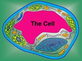
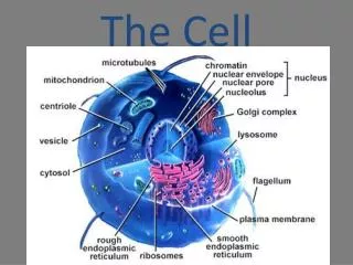
An eukaryotic cell has internal membranes, which partition the cell into compartments . These membranes also participate in metabolism as many enzymes are built into membranes. The general structure of a biological membrane is a double layer of phospholipids and diverse proteins. Each type of membrane has a unique combination of lipids and proteins for its specific functions. For example, those in the membranes of mitochondria function in cellular respiration. Internal membranes compartmentalize the functions of a eukaryotic cell
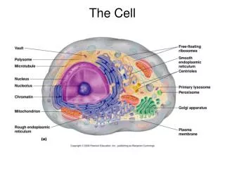
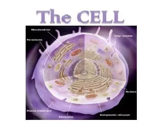
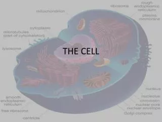
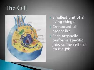
B- Eukaryotic Cell Eu: True Karyon: Nucleus Plant Cell Animal Cell What are the functions of cell organelles ? Compare between Animal and Plant cell?
Euokaryotes: Euo = true karyot = nucleus . Plant and animals have real nucleus, surrounded with nuclear membrane. • -The Bacteria and the virus’s have no real nucleus they contain nucleiod region (no nuclear membrane) were the very simple genetic material (DNA or chromosome) • -The prokaryotic cells (bacteria and viruses) also have a very simples cell structure cell wall, cell membrane, cytoplasm, ribosome’s and nucleiod area for a very simple genetic material (DNA or RNA) and cilia or flagella. • The euokayotic cells have a very complex structure and many cell organelles (Look at the book page 112. 6th ed.)
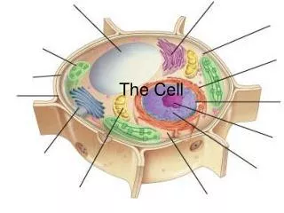
المادة الوراثية الشبكة الإندوبلازمية نوية النواة الجدار النووى سوط حركى جسم مركزى ريبوسوم حهاز جولـﭽـى غشاء بلازمى حلمات دقيقة ميتوكوندريا جسم مُحلل الهيكل الخلوى
فجوة مركزية بلاستيدة خضراء الجدار الخلوى ثقوب بينية
Cytokinesis in animal cell: • Cytokinesis, division of the cytoplasm, typically follows mitosis. • In animals, the first sign of cytokinesis (cleavage) is the appearance of a cleavage furrow in the cell surface near the old metaphase plate.
Cytokinesis in plants, which have cell walls, involves a completely different mechanism. • During telophase, vesicles from the Golgi coalesce at the metaphase plate, forming a cell plate. • The plate enlarges until its membranes fuse with the plasma membrane at the perimeter, with the contents of the vesicles forming new wall material in between.
Prokaryotic vs. eukaryotic gene structure prokaryotes: polycistronic transcripts eukaryotes: monocistronic transcripts