Download
1 / 38
410 likes | 476 Views
Venous Thrombosis in Pregnancy. Dr. R. sami Pulmonologist IUMS. VTE in pregnancy. Pregnant women are at an increased risk for venous thromboembolic disease (VTE) 1 in 1000 pregnancies 2-4 fold increase compared to non-pregnant state Cesarian delivery > vaginal delivery

E N D
Venous Thrombosis in Pregnancy Dr. R. sami Pulmonologist IUMS
VTE in pregnancy • Pregnant women are at an increased risk for venous thromboembolic disease (VTE) • 1 in 1000 pregnancies • 2-4 fold increase compared to non-pregnant state • Cesarian delivery > vaginal delivery • 2/3 of DVT occur antepartum • 43-60% of PE occur 4-6 weeks after delivery • PE is the major non-obstetric cause of maternal mortality • 2/100 000 pregnancies
Why is the risk greater in pregnancy? • Pathopsysiology • Increased venous capacity (estrogen) • Increased plasma volume • Compression of IVC • Increased levels of coagulation factors (fibrinogen, factor VII) • Decreased levels of natural anticoagulants (protein S) • Acquired protein C resistance
Independent risk factors • Bed rest • Multiparity • Advanced maternal age (>35 yo) • Overweight • Personal or family history of VTE • Preeclampsia
خانم 28 ساله باردار 14 هفته با درد و تورم ساق پای چپ از 2 روز قبل مراجعه کرده است: تشخیص افتراقی ؟
Ruptured Baker's cyst • Erysipelas
Malignancy Surgery Trauma Pregnancy Oral contraceptives or hormonal therapy Immobilization Inherited thrombophillia Presence of venous catheter Congestive failure Antiphospholipid antibody syndrome Hyperviscosity Nephrotic syndrome Inflammatory bowel disease DVT – VTE Risk Factors
Cancer Paralysis or plaster immobilization Bedrest > 3d or surgery in past 4 wks Localized tenderness Entire leg swollen Calf > 3cm larger than unaffected leg Pitting edema greater than unaffected leg Collateral superficial veins DVT – Wells Score The following were assigned a point value of 1 if present: • Alternative diagnosis more likely than DVT = - 2 points • Probability High (≥ 3), Moderate (1-2) or Low (0 or less) • DVT risk: High – 75%, Moderate – 17%, Low – 3% Wells PS, Andersen DR, Bormanis J et al. Lancet. 1997;350:1795-8
DVT – D-Dimer • Fibrin degradation product elevated in active thrombosis • Negative ….. Positive…. • Preferred test • Quantitative Rapid ELISA – sensitivity 96/95% for DVT/PE • Other methods include latex agglutination and RBC agglutination (SimpliRED) Stein PD, Hull RD, Patel KC, et al. Ann Int Med. 2004;140(8):589-602
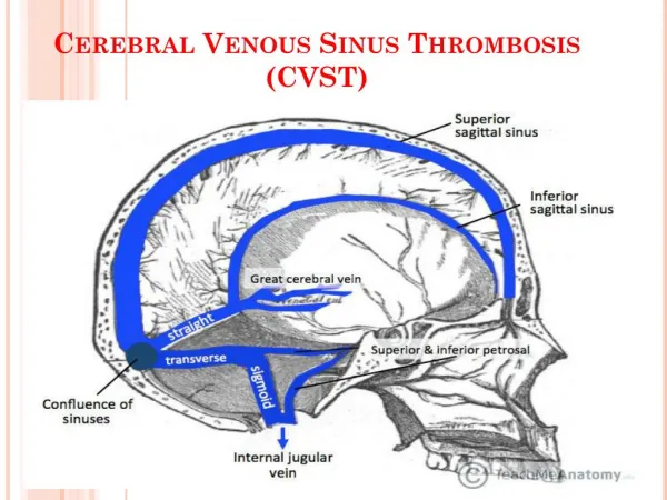
90% of DVT during pregnancy occurs on the left side. • A significant proportion of DVT in pregnancy occurs in the pelvic veins and therefore, may not be picked up by routine testing.
Calf • Proximal • Ultrasonography ???? • Approach ????
خانم 24 ساله باردار 32 هفته با تنگی نفس از 3 روز قبل مراجعه کرده است : • تشخیص افتراقی؟؟ بیمار مبتلا به آمبولی ریه با چه علایمی مراجعه می کند؟
signs and symptoms • Clinical signs and symptoms of PE are rarely encountered together; the classic symptoms are as follows: • Dyspnea - 82% • Abrupt onset of chest pain - 49% • Cough - 20% • The most common presenting signs of PE are as follows: • Tachypnea • Crackles • Tachycardia
Patients with massive PE may present with the following: • Syncope • Hypotension • Pulseless cardiac electrical activity • Death
LEFt • L- Symptoms in the left lower extremity • E-Edema: • Mid-calf circumference difference of ≥ 2cm • Ft- First trimester presentation • No finding 0 • 1 finding 16% • 2 or 3 findings 58%.
Para clinic • If DVT or PE is suspected, the patient should begin anticoagulation treatment until further investigation excludes VTE. • ECG • ABG • CXR • CT • Scan
Abnormal V/Q Scan Perfusion Ventilation
fetal risks from radiation doses of less than 50 mGy are negligible • doses of 100 mGy and more result in a combined increased risk of organ malformation and the development of childhood cancer of only about 1%
even a combination of imagings( chest radiography, lung scintigraphy, CT pulmonary angiography, and traditional pulmonary angiography )exposesthe fetus to around 1.5 mGy of radiation(below the accepted limit of 50 mGy)
Fetal dose by CTPA is about 0.03-0.66 mGy • lung scintigraphy is more (about 0.32-0.74 mGy) • scintigraphy, radiotracer is injected intravenously and lead to direct fetal exposure • estimated breast dose from CTPA is 150 times more than scintigraphy
risks of iodine contrast agents are similar to general population • no fetal risks from intravenous contrast (they are classified as category B by FDA) • infant thyroid function