Download

1 / 65
820 likes | 2.55k Views
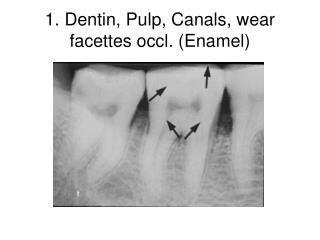
Structure and Functions of the Dentin-Pulp Complex. Husain Keylani R1 endo board. Dentin-pulp complex ? E mbryologically , histologically and functionally; dentin and pulp should be considered the same. This entity is exemplified by the classic functions of the pulp.

E N D
Structure and Functions of the Dentin-Pulp Complex Husain Keylani R1 endo board
Dentin-pulp complex ? • Embryologically, histologically and functionally; dentin and pulp should be considered the same. This entity is exemplified by the classic functions of the pulp. • The transition to pulp occurs with the initiation of dentin formation.
The pulp • The pulp incompressible, the total volume within the pulp chamber cannot be greatly increased. • Thus Inflammatory reaction results in an increase in pulp tissue pressure instead of volume. (Heyeraas et al,1992)
Development • The Primary oral cavity or stomodeum(lined with ectoderm )is a depression between the brain and the pericardium in an embryo, • It is separated from the upper end of the foregut by (bucco-pharyngeal membrane ,27 days) Mid-brain bucco-pharyngeal membrane stomodeum pericardium
Development • Most of the C.T. cells underlying the oral ectoderm are neural crest cells or ectomesenchyme in origin
Development • Neural crest travel down the sides of the head into the maxilla and mandible to form the toothgerm
Development During sixth week ,tooth formation begin as localized proliferation of ectoderm to form two horseshoe-shaped structures called primary epithelial band or primary dental laminaewhich split into vestibular & dental lamina Oral vestibule Dental organ &
Stage of development • Formation of the teeth is a continuous process has been divided in to three stages : • bud stage • Cap stage • bell stage
Cont. • 1-The bud stage: • the initial stage , • dental lamina proliferate • to the adjacent ectomesenchyme
Cont. 2- Cap stage : the dental lamina cont. proliferate- aconcavity. • The outer cells cuboidal and constitute the outer enamel epithelium . • The inner cells elongated and represented the inner enamel epithelium . Between them is network of cells termed (the stellate reticulum)
Cont. • 3-bellstage (18): • The last period of growth is also known as histodifferentiation • In the cervical loop where the outer and the inner enamel epithelia are joined continue to proliferate . • The tooth development enters the bell stage • During (BV) become established in the dental papilla
Cell differentiation mechanisms • Embryonic development of any tissue is prompted by interaction with an adjacent tissue. • Complex epithelial and mesenchymal interactions occur which direct the ameloblasts and odontoblasts diff. by changes in gene expression.
Cont. • Epithelial- ectomesenchymal inductive interaction during normal odontogenesis lead to: 1- Cytodifferentiation of dentin and enamel forming cells. 2-dental hard tissue formation
Mechanisms of interaction A: cell-to-cell interaction 1-Direct cell-to-cell contact. 2-The transmission of molecules synthesized and secreted by one cell and then captured by surface receptors of another cells. • CAMs(cell adhesion molecule )mediate morphogenesis. through contrall cell proliferation • Cell contian membrane protein called (integrin)receptor for CAMs. integrin CAMs
Cont… B:Cell-to-ECM interaction. • Carry by substrate adhesion molecule (SAMs) the best study of the ECM are fibronectins. • G.factors are polypeptides produced by cells that initiate proliferation, migration and diff. of a variety of cells.
Cont… • Dental basement membrane: • It exists between IDE(inner dental epithelium and the underneath dental mesenchyme • It consist of (thin basal lamina & layer of ECM)
Cont… • Dental basement membrane: • It exists between IDE(inner dental epithelium and the underneath dental mesenchyme • It consist of (thin basal lamina & layer of ECM) • BL is compsed of type IV collagen (as areceptors) whichhas binding sites for other BM consituents. (like laminin ,fibrinoctin ,proteoglycan) • Laminin is the CAM in the basement membrane
Cont… • Odontoblast cell surface proteoglycans act as receptors for matrix molecules .signals from matrix components influence migration and diff. of odontoblasts.
Differentiation of Odontoblasts • occurs during the bell stage. • Preameloblasts diff. at a faster rate than odontoblasts, but dentin matrix is formed before enamel matrix. • There is still mitotic activity among the relatively immature cells of the inner enamel epithelium., mitotic activity ceases and the cells elongate.
Cont.Differentiation of Odontoblasts • With the onset of differentiationa single layer of cell(preodontoblasts) align themselves along the basement membrane separating the inner enamel epithelium from the dental papilla. IEE PO BM DP
Cont.Differentiation of Odontoblasts • These cells stop dividing and elongate into short columnar cells with basally situated nuclei • Several cytoplasmic projections from each of these cells extend toward the basal lamina.
Cont.Differentiation of Odontoblasts • As the odontoblasts continue to differentiate, they become progressively more elongated • And cytoplasmic processes from these cells extend through the BM toward the basal lamina, and more and more collagen fibrils appear within the ECM.
Cont.Differentiation of Odontoblasts • More odontoblastic diff elongation with characteristic of protein-secreting cells more collagen fibrils appear in the ECM and more defined large odontoblasticprocess toward BL.
Cont.Differentiation of Odontoblasts • Theodontoblastsreach full maturity and become tall columnar cells • Production of the initial dentin matrix involves the formation, organization, and maturation of collagen fibrils and proteoglycans. • As more collagen fibrils accumulate subjacent to the basal lamina,(The lamina becomes discontinuous and eventually disappears. )
Cont.Differentiation of Odontoblasts • The odontoblasts reach full maturity and become tall columnar cells • Production of the initial dentin matrix involves the formation, organization, and maturation of collagen fibrils and proteoglycans. • As more collagen fibrils accumulate subjacent to the basal lamina,(The lamina becomes discontinuous and eventually disappears. ) • Some of Odon. processes extend toward the ameloblasts • Some of these become interposed between the processes of ameloblasts, resulting in the formation of enamel spindles
Cont.Differentiation of Odontoblasts • At the onset of dentinogenesis the dental papilla becomes the dental pulp • As predentin matrix is formed, the odontoblasts commence to move toward the central pulp, depositing matrix. • Within this matrix a process from each odontoblast becomes accentuated and remains to form the primary odontoblast process. • It is around these processes that the dentinal tubules are formed.
Root • Develop after completion of enamel formation. • Proliferateion (inner and outer enamel epithelia), • and form a structure known as the Hertwig epithelial root sheath (HERS) • (HERS) determine the size and shape of root of the tooth .
Root • The first layer of the dentin matrix mineralizes gap appear in the root sheath allowing mesenchymal cells from the dental sac to move into contact with the newly formed dentine . • These cells then differntiation into cementoblast and deposit cementum matrix on the root dentine.
Epithelial Rests of Malassez • Persistant of epithelial cells of the root sheath in the PDL after tooth development. • Some of them retain & under go division • Later produce periapical cyst. (Trowbridge,1967)
Accessory Canals • During root formation : • a break develops in the continuity of the sheath , producing a small gap ,dentinogenesis does not take place opposite the defect . • The result is small “accessory “ canal between the dental sac and the pulp . • If periodontal tissue lose their integrity , m/o go pulp
DENTIN • Composition of fully mature dentin : • INORGANIC MATERIAL • ORGANIC MALTERIAL WATER • 20% • ~91%(collagen) I,V. • ~9%(non-collagen) • *phosphoproteins • *proteoglycans • *Gla proteins • *acidic glycoproteins • *Gr. Factors+lipids • 70% • Hydroxyapatite 10%
Types of the dentin 1-Developmental dentin : is forms during tooth developmet &classified as: A-orthodentin: - the tubular form of dentin
Cont. Types of the dentin B-mantle dentin -is the first formed dentin and is situated immediately subjacent to the enamel or cementum. -( 150μm, less mineralized and softer). (Herr P et al,1986)
Cont. Types of the dentin C-Circumpulpal dentin : - it constitutes the major part of developmental dentin. - oriented at right angles to the long axis of the dentinal tubules. -500nm in diameter
Cont. Types of the dentin • 2-Secondary dentin: -Regular dentin which forms phisiologically after the root is fully developed. A-Primary physiological dentin B-Secondary physiological dentin
Cont. Types of the dentin • 3-Tertiary dentin: -Irregular dentin forms in response to abnormal stimuli • Reparative D filling • Reactionary D irritant
Predentin *Is unmineralized organic matrix of dentin between the odontoblast layer and the mineralized dentin • type I , II collagens. • Noncollagenous elements (proteoglycans,Phosphophoryn) • growth factors (Roberts-Clark D,2000) Dentin Predentin Odontoblasts Pulp cells
Mineralization • Caph.accomu. In vesicle (prd) grow • Rupsure release mix • the mix adjoin to form advance crystl merge to form small globules • The globule expand and fuse with other untill become mineraized • Cont. in growing and increase mineral content. A B E C D Mineralization
Cont.Mineralization Mineralized dentin Predentin Mineralization front Odontoblasts
Dentinal tubules • Dentinal tubules occupy from 1% (superficial dentin) to 30% (deep dentin) of the volume of intact dentin. (Garberoglio,Brannstrom 1976) • It slightly tapered , wider portion toward the pulp.(no pretubular d)
S shape DEJ- pulp. b/c (crowding of odontoblasts) • It converge ? the surface of the pulp chamber has a much smaller area than the surface of dentin along the DEJ. This results in a progressive increase in dentin permeability Coronal dentin Cont.Dentinal tubules
Cont.Dentinal tubules • Near the DEJ the dentinal tubules ramify into one or more terminal branches.
Peritubular and intertubular dentin • Peritubular Dentin: is dentin lining the tubule , whereas that between the tubules is known as intertubular dentin • minir+hard • < collagn
Dentinal Sclerosis • Partial or complete obturation of dentinal tubules. as result of aging or develop in response to stimuli such as attrition or caries • It helps the pulp from irritation
Cont.Dentinal Sclerosis It is either: Physiologic:acceleration of peritubular dentin formation in the apical third of the root. Pathologic:Dentinal tubules are blocked byhydroxyapatite and whitlockite crystals (carious dentin, attrited dentin)