Download
1 / 95
990 likes | 1.44k Views
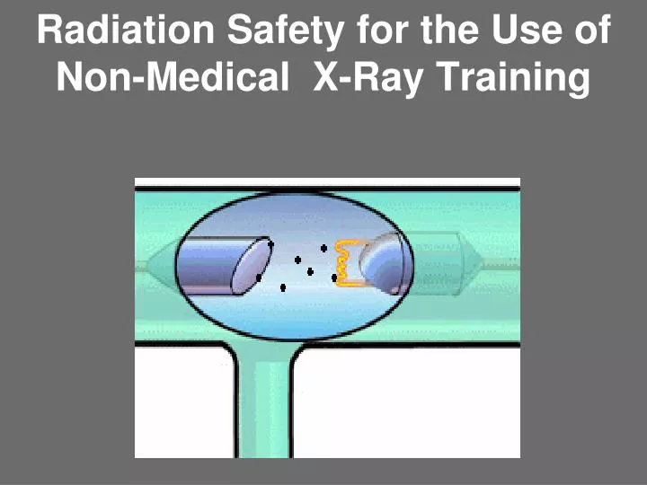
Radiation Safety for the Use of Non-Medical X-Ray Training. Instructor. Dennis Widner Health Physicist – Training Radiation Safety Office University of Georgia 706-542-0526. INTRODUCTION.

E N D
Instructor Dennis Widner Health Physicist – TrainingRadiation Safety OfficeUniversity of Georgia 706-542-0526
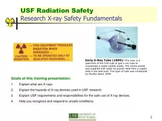
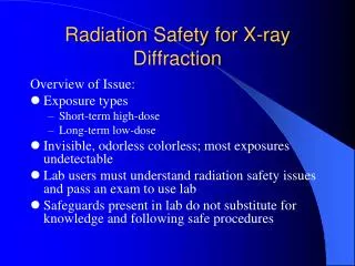
INTRODUCTION The purpose of this safety presentation is to increase your knowledge in order to enable you to perform your job safely by adhering to proper radiation protection practices while working with or around x-ray-generating devices. This course will inform you about the policies and procedures you should follow to reduce the risk of exposure to the ionizing radiation produced by x-ray-generating devices.
Georgia DHR Training Outline • Fundamentals of Radiation Safety • Characteristics of radiation • Units of radiation measurement • Significance of radiation dose and exposure (radiation protection standards and biological effects) • Sources and levels of radiation • Methods of controlling radiation dose (time, distance, and shielding) • Radiation Detection Instrumentation to be Used • Use of radiation survey instruments (operation, calibration, limitations) • Use of personnel monitoring equipment (dosimetry) • Radiographic Equipment to be Used • Remote handling equipment • Radiographic exposure devices • Operation and control of x-ray equipment • Pertinent Federal and State Regulations • The Registered Users Written Operating and Emergency Procedures • Case Histories of Radiography Accidents
INSTRUCTION OF PERSONNEL • The registrant (UGA) shall assure that all radiation machines and associated equipment under his control are operated only by individuals instructed in safe operating procedures and are competent in the safe use of the equipment. • UGA shall also assure that persons operating radiation machines and associated equipment receive 2 hours of training in radiation safety within 90 days after employment. Training can be performed by the researcher or by attending the UGA/RSO X-ray class.
TRAINING DOCUMENTATION • Each user of a radiation machine and associated equipment must have documented training records for operation and safety. These records shall be maintained in the laboratory for the lifetime of operation. • The Researcher, Principal Investigator or Supervisor is responsible for these records.
ORGANIZE RECORDS IN 3 RING BINDER MAINTENANCE INSPECTIONS OPERATION PROCEDURE EMERGENCY PROCEDURE QUARTERLY CHECKS TRAINING REGISTRATION EQUIPMENT • Equipment Specifications and output • Registration documents • Training Records • Quarterly Safety Checks/ Surveys • Operation and Emergency Procedures • Maintenance records • Inspection Results
Compentency • Identification of the radiation hazards associated with the operation of your equipment. • Understanding the significance of equipment warnings, safety devices and interlocks. • Adherence to operating procedures • Recognize acute exposure symptoms andhow to report an acute exposure • Any exposure should be reported to UGA Radiation Safety Office at 542-5801 or 542-0107
How X-Ray Machines Work Tube Leakage Vacuum Tube Cathode - Electrons Anode - Target (W) (Al)(Mo) Wire Filament Filter Power
INTENTIONAL An intentional x-ray device is designed to generate an x-ray beam for a particular use. Intentional x-rays are typically housed within a fixed, interlocked and/or shielded enclosure or room. Examples include x-ray diffraction and fluorescence analysis systems, flash x-ray systems, medical x-ray machines, and industrial cabinet and non-cabinet x-ray installations. INCIDENTAL An incidental x-ray device produces x-rays that are not wanted or used as a part of the designed purpose of the machine. Examples of incidental systems are computer monitors, televisions, electron microscopes, high-voltage electron guns, electron-beam welding machines, and electrostatic separators.
Intentional Analytical X-Ray Devices Analytical X-Ray Devices Analytical x-ray devices use x-rays for diffraction or fluorescence experiments as research tools, especially in materials science. ANSI N43.2 defines two types of analytical x-ray systems: enclosed beam and open beam.
Safety requirements and features for analytical systems include the following: · control panel labels with the words “CAUTION — HIGH INTENSITY X-RAY BEAM · fail-safe lights with the words “X-RAYS ON” near x-ray tube housings, · fail-safe indicators with the words “SHUTTER OPEN” for beam shutters,
·fail-safe interlocks on access doors and panels, beam stops or other barriers, and appropriate shielding.
Enclosed-Beam System In an enclosed-beam system, all possible x-ray paths (primary and diffracted) are completely enclosed so that no part of a human body can be exposed to the beam during normal operation. Because it is safer, the enclosed-beam system should be selected over the open-beam system whenever possible. The x-ray tube, sample, detector, and analyzing crystal (if used) must be enclosed in a chamber or coupled chambers. The sample chamber must have a shutter or a fail-safe interlock so that no part of the body can enter the chamber during normal operation.
The dose rate measured at 10 inches (25 cm) from the apparatus must not exceed 2.0 mR per hour during normal operation.
Open-Beam System In an open-beam system, one or more x-ray beams are not enclosed, making exposure of human body parts possible during normal operation. The open-beam system is acceptable for use only if an enclosed-beam system is impractical for any of the following reasons: · a need for making adjustments with the x-ray beam energized, · a need for frequent changes of attachments and configurations, · motion of specimen and detector over wide angular limits, or · the examination of large or bulky samples.
An open-beam x-ray system must have a guard or interlock to prevent entry of any part of the body into the primary beam. Each port of the x-ray tube housing must have a beam shutter with a conspicuous shutter-open indicator of fail-safe design. The dose rate from tube leakage at 2 inches (5 cm) from the surface of the tube housing must not exceed 25 mR per hour during normal operation. The dose rate at 2 inches (5 cm) from the surface of the HV power supply must not exceed 0.5 mR per hour during normal operation.
NON-MEDICAL FLUOROSCOPY • “Hand –held” fluoroscopes shall not be used • The dose rate due to transmission through the image receptor • shall not exceed 2 mR/hr at 4 inches ( 10 cm) from any point • On the receptor. • The maximum x-ray dose shall not exceed 0.5 mR in any one • hour measures at 2 inches ( 5 cm) from any readily accessible • machine surface
Intentional Industrial X-Ray Devices Industrial x-ray devices are used for radiography; for example, to take pictures of the inside of an object as in a medical chest x-ray or to measure the thickness of material. ANSI N43.3 defines three classes of industrial x-ray installations: • Cabinet • exempt shielded • shielded.
Incidental X-Ray Devices In a research environment, many devices produce incidental x-rays. Any device that combines high voltage, a vacuum, and a source of electrons could, in principle, produce x-rays. For example, a television or computer monitor generates incidental x-rays, but in modern designs the intensity is low, much less than 0.5 mR per hour. Occasionally, the hazard associated with the production of incidental x-rays is recognized only after the device has operated for some time. If you suspect an x-ray hazard, contact UGA Radiation Safety to survey the device.
Electron Microscopes The exposure rate during any phase of operation of an electron microscope at the maximum rated continuous beam current for the maximum rated accelerating potential should not exceed 0.5 mR per hour at 2 inches (5 cm) from any accessible external surface.
X-Ray Safety Training Fundamentals of Radiation Safety
Characteristics of Radiation What is Radioactivity? What is scatter ? What are X-rays ?
Radioactivity Definition Any spontaneous change in the state of a nucleus accompanied by the release of energy. Major Types alpha () particle emission beta () particle emission gamma () decayX-ray (X) Characteristic and Bremsstrahlung
X-Rays X-rays are photons (electromagnetic radiation) which originate in the energy shells of an atom, as opposed to gamma rays, which are produced in the nucleus of an atom.
Soft vs. Hard X-rays X-rays from about 0.12 to 12 keV (10 to 0.10 nm wavelength) are classified as "soft" X-rays, and from about 12 to 120 keV (0.10 to 0.01 nm wavelength) as "hard" X-rays, due to their penetrating abilities.[3]
X-ray Tube Target Material In medical X-ray tubes the target is usually Tungsten (W) or a more crack-resistant alloy of Rhenium (Re) (5%) and tungsten (95%), but sometimes Molybdenum (Mo) for more specialized applications, such as when soft X-rays are needed as in mammography. In crystallography, a Copper (Cu) target is most common, with Cobalt (Co) often being used when fluorescence from Iron (Fe)content in the sample might otherwise present a problem.
X-Ray Scatter When x-rays pass through any material, some will be transmitted, some will be absorbed, and some will scatter. The proportions depend on the photon energy and the type of material. X-rays can scatter off a target to the surrounding area, off a wall and into an adjacent room, and over and around shielding. A common mistake is to install thick shielding walls around an x-ray source but ignore the need for a roof, based on the assumption that x-rays travel in a straight line. The x-rays that scatter over and around shielding walls are known as skyshine.
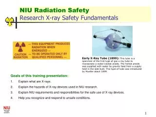
On November 8, 1895, at the University of Wurzburg, Wilhelm Roentgen's attention was drawn to a glowing fluorescent screen on a nearby table. Roentgen immediately determined that the fluorescence was caused by invisible rays originating from the partially evacuated glass Hittorf-Crookes tube he was using to study cathode rays (i.e., electrons). Surprisingly, these mysterious rays penetrated the opaque black paper wrapped around the tube. Roentgen haddiscovered X rays, a momentous event that instantly revolutionized the field of physics and medicine. Wilhelm Conrad Roentgen (1845-1923)
X-rays X-rays are Electromagnetic Radiation (EMR)
EM radiation can be viewed as a waves or bundles ofenergy called photons. Electromagnetic radiation (EM) is the transport of energy through space as a combination of electric and magnetic fields The EM wave can be visualized as an oscillating electric field with a similar varying magnetic field changing with time and at right angles to it.
X-ray wavelengths are typically measured in units of angstroms Å = 1 E-10 meters(0.0000000001 m)1 nanometer = 1 E-9 meter 1 nm = 10Å The x-ray region is normally considered to be that region of the EM spectrum lying between 0.1 and 100 Åin wavelengthorX-ray energies between 0.1 and 100 keV
How small is an angstrom? • The point of a needle is about 1 million angstroms in diameter. • Fingernails grow at about 50 angstroms per second. • One angstrom is to a grain of sand, as a child's wading pool is to the Atlantic Ocean.
Types of X-rays Characteristic vsBremsstrahlung X-rays can be produced by either by the interaction of the bombarding electrons that are braked by the Coulomb force field of the target nuclei (Bremsstrahlung x-ray production) Collision interactions with atomic electrons of the target material (characteristic x-ray emission).
Ionizing Radiation Definition - Any type of radiation possessing enough energy to eject an electron from an atom, thus producing an ion. X-Rays and Gamma photons are both electromagnetic radiations that have the energy to ionize atoms X-Ray
Fundamentals of Radiation Safety Units of Radiation Measurement
DOSE UNITS OF MEASURE • Ionizing radiation is measured in the following units: • · roentgen (R), the measure of exposure to radiation, defined by the ionization caused by x-rays in air. • · rad, the radiation absorbed dose or energy absorbed per unit mass of a specified absorber. • · rem, the roentgen equivalent man or dose equivalent.
Georgia Radiation Dose Units MilliRoentgen (mR)or Roentgen (R) Georgia Radiation Dose rate Units MilliRoentgens per hour (mR/hr)or Roentgens per hour (R/hr)
Fundamentals of Radiation Safety Significance of Radiation Dose and Exposure
Health Effects of Radiation Ionizing Radiation can directly and indirectly damage DNA Acute Exposure Effects (Stochastic) Radiation in large doses in a short time causes observable damage ….observable at >25 Rem Chronic Exposure Effects (Non-stochastic) The effects from radiation exposure decrease as the dose rate is lowered. Spreading the dose over a longer period reduces the effects. Much of the controversy over radiation exposure centers on the question of how much damage is done by radiation delivered at low doses or low dose rates. DNA Double Helix Radiation
Dose Response Model Known Effects Atomic Bomb Survivors Uranium Miners Radium Dial Painters Medical Patients Health Effect (cancer) • Linear No Threshold Dose Curve • Decreased Health Effects Theory • Threshold Dose Theory • Increased Health Effects Theory 4 Theo. Debated Effects 1 2 The NRC and The State of Georgia Follow the Linear No Threshold Theory 3 Dose (rem)
How does radiation cause health effects? Radioactive materials that decay spontaneously produce ionizing radiation, which has sufficient energy to strip away electrons from atoms (creating two charged ions) or to break some chemical bonds. Any living tissue in the human body can be damaged by ionizing radiation. The body attempts to repair the damage, but sometimes the damage is too severe or widespread, or mistakes are made in the natural repair process. The most common forms of ionizing radiation are alphaand beta particles, or gamma and X-rays.
What kinds of health effects occur from exposure to X-rays? In general, the amount and duration of x-ray exposure affects the severity or type of health effect. There are two broad categories of health effects: stochastic and non-stochastic.
Stochastic effects are associated with long-term, low-level (chronic) exposure to radiation. ("Stochastic" refers to the likelihood that something will happen.) Increased levels of exposure make these health effects more likely to occur, but do not influence the type or severity of the effect. Stochastic Health Effects Cancer is considered by most people the primary health effect from radiation exposure. Simply put, cancer is the uncontrolled growth of cells. Ordinarily, natural processes control the rate at which cells grow and replace themselves. They also control the body's processes for repairing or replacing damaged tissue. Damage occurring at the cellular or molecular level, can disrupt the control processes, permitting the uncontrolled growth of cells--cancer. This is why ionizing radiation's ability to break chemical bonds in atoms and molecules makes it such a potent carcinogen. Other stochastic effects also occur. Radiation can cause changes in DNA, the "blueprints" that ensure cell repair and replacement produces a perfect copy of the original cell. Changes in DNA are called mutations. Sometimes the body fails to repair these mutations or even creates mutations during repair. The mutations can be teratogenic or genetic. Teratogenic mutations affect only the individual who was exposed. Genetic mutations are passed on to offspring.
Non-Stochastic Health Effects Non-stochastic effects appear in cases of exposure to high levels of radiation, and become more severe as the exposure increases. Short-term, high-level exposure is referred to as 'acute' exposure. Many non-cancerous health effects of radiation are non-stochastic. Unlike cancer, health effects from 'acute' exposure to radiation usually appear quickly. Acute health effects include burns and radiation sickness. Radiation sickness is also called 'radiation poisoning.' It can cause premature aging or even death. If the dose is fatal, death usually occurs within two months. The symptoms of radiation sickness include: nausea, weakness, hair loss, skin burns or diminished organ function. Medical patients receiving radiation treatments often experience acute effects, because they are receiving relatively high "bursts" of radiation during treatment.
What is the cancer risk from radiation? How does it compare to the risk of cancer from other sources? To give you an idea of the usual rate of exposure, most people receive about 3 tenths of a rem (300 mrem) every year from natural background sources of radiation (mostly radon). Each radionuclide represents a somewhat different health risk. However, health physicists currently estimate that overall, if each person in a group of 10,000 people exposed to 1 rem of ionizing radiation, in small doses over a life time, we would expect 5 or 6 more people to die of cancer than would otherwise. ( 0.06%) In this group of 10,000 people, we can expect about 2,000 to die of cancer from all non-radiation causes. The accumulated exposure to 1 rem of radiation, would increase that number to about 2005 or 2006.