Download
1 / 45
750 likes | 1.6k Views
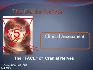
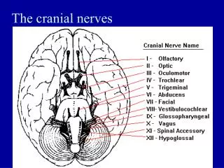
The Cranial Nerves. SHANDONG UNIVERSITY Liu Zhiyu. Names of Cranial Nerves. Ⅰ Olfactory nerve Ⅱ Optic nerve Ⅲ Oculomotor nerve Ⅳ Trochlear nerve Ⅴ Trigeminal nerve Ⅵ Abducent nerve Ⅶ Facial nerve Ⅷ Vestibulocochlear nerve Ⅸ Glossopharyngeal nerve

E N D
The Cranial Nerves SHANDONG UNIVERSITY Liu Zhiyu
Names of Cranial Nerves • Ⅰ Olfactory nerve • Ⅱ Optic nerve • Ⅲ Oculomotor nerve • Ⅳ Trochlear nerve • Ⅴ Trigeminal nerve • Ⅵ Abducent nerve • Ⅶ Facial nerve • Ⅷ Vestibulocochlear nerve • Ⅸ Glossopharyngeal nerve • Ⅹ Vagus nerve • Ⅺ Accessory nerve • Ⅻ Hypoglossal nerve
I Olfactory (oh) II Optic (oh) III Oculomotor (oh) IV Trochlear (to) V Trigeminal (1-3) (touch) VI Abducens (and) VII Facial (feel) VIII Vestibulocochlear (very) IX Glossopharyngeal (good) X Vagus (velvet) XI Accessory (ah) XII Hypoglossal (heaven)
How to Remember CN I-XII Oh! Oh! Oh! To Touch And Feel Very Good Velvet! Ah Heaven!
Functional Components • General somatic afferent fibers (GSA): transmit exteroceptive and proprioceptive impulses from head and face to somatic sensory nuclei • Special somatic afferent fibers (SSA): transmit sensory impulses from special sense organs of vision, equilibrium and hearing to the brain • General visceral afferent fibers (GVA): transmit interoceptive impulses from the viscera to the visceral sensory nuclei • Special visceral afferent fibers (SVA): transmit sensory impulses from special sense organs of smell and taste to the brain
Functional Components • General somatic efferent fibers (GSE): innervate skeletal muscles of eye and tongue • Special visceral efferent fibers (SVE): transmit motor impulses from the brain to skeletal muscles derived from brachial (gill) arches of embryo. These include the muscles of mastication, facial expression and swallowing • General visceral efferent fibers (GVE): transmit motor impulses from the general visceral motor nuclei and relayed in parasympathetic ganglions. The postganglionic fibers supply cardiac muscles,smooth muscles and glands
Classification of Cranial Nerves • Sensory cranial nerves: are composed entirely of afferent (sensory) nerve fibers bringing sensations the brain • Ⅰ Olfactory nerve • Ⅱ Optic nerve • Ⅷ Vestibulocochlear nerve • Motor cranial nerves: are composed entirely of efferent (motor) fibers • Ⅲ Oculomotor nerve • Ⅳ Trochlear nerve • Ⅵ Abducent nerve • Ⅺ Accessory nerve • Ⅻ Hypoglossal nerve • Mixed cranial nerves: possess both sensory and motor fibers--- • Ⅴ Trigeminal nerve, • Ⅶ Facial nerve, • Ⅸ Glossopharyngeal nerve • Ⅹ Vagus nerve
Olfactory Nerve Olfactory cells (SVA)→ Cribriform foramina → Olfactory bulb
Optic Nerve Ganglion cells (SSA) → Optic canal → Lateral geniculate body
Vestibulocochlear Nerve Vestibular ganglion(SSA)↘↗Vestibular nuclei Internal acoustic meatus Cochlear ganglion (SSA)↗↘Cochlear nuclei
Motor Cranial Nerves Superior orbital fissure Ⅲ Ⅳ Ⅵ Hypoglossal canal Ⅻ Ⅹ Jugular foramen Ⅺ
Oculomotor Nerve • Components • General somatic efferent fibers (GSE) • General visceral efferent fibers (GVE) • Main action-supplies • Superior, inferior and medial recti; inferior obliquus; levator palpebrae superioris • Sphincter pupillea and ciliary muscle • Ciliary ganglion : lies between optic nerve and lateral rectus Oculomotor nerve
Abducent nerve Abducent Nerve
Trochlear n. Oculomotor n. Abducent n.
Accessory Nerve • Cranial roots • Originate from nucleus ambiguus • Join the spinal roots to exit the jugular foramen • Join the vagus nerve and distribute to the muscles of pharynx and larynx • Spinal roots • Originate from nucleus of accessory nerve • Ascend through the foramen magnum and exit the cranium through the jugular foramen • Innervate the sternocleidomastoid and trapezius muscles
Hypoglossal Nerve Nucleus of hypoglossal nerve( GSE) → Hypoglossal canal → Muscles of tongue Hypoglossal nerve
Trigeminal Nerve (Ⅴ) Components of fibers • SVE fibers: originate from motor nucleus of trigeminal nerve, and supply masticatory muscles • GSA fibers: transmit facial sensation to sensory nuclei of trigeminal nerve, the GSA fibers have their cell bodies in trigeminal ganglion, which lies on the apex of petrous part of temporal bone
Trigeminal Nerve (Ⅴ) Ophthalmic nerve(Ⅴ1, sensory) • Leave the skull through the superior orbital fissure, to enter orbital cavity • Branches • Frontal nerve • Supratrochlear nerve • Supraorbital nerve • Lacrimal nerve • Nasociliary nerve
Trigeminal Nerve (Ⅴ) Ophthalmic nerve • Branches • Frontal nerve • Lacrimal nerve • Nasociliary nerve • Distribution • Sensation from cerebral dura mater • Visual organ • Mucosa of nose • Skin above the eye and back of nose
Trigeminal Nerve (Ⅴ) Maxillary nerve(Ⅴ2, sensory) • Leave skull through foramen rotundum • Branches • Infraorbital nerve • Superior alveolar nerve • Zygomatic nerve • Pterygopalatine nerve
Trigeminal Nerve (Ⅴ) Maxillary nerve • Branches • Infraorbital nerve • Superior alveolar nerve • Zygomatic nerve • Pterygopalatine nerve • Distribution • Sensation from cerebral dura mater • Maxillary teeth and gum • Mucosa of nose, mouth and maxillary sinus • Skin between eye and mouth
Trigeminal Nerve (Ⅴ) Mandibular nerve(Ⅴ3, mixed) • Leave the skull through the foramen ovale to enter the infratemporal fossa • Branches • Auriculotemporal nerve • Buccal nerve • Lingual nerve • Inferior alveolar nerve • Nerve of masticatory muscles
Trigeminal Nerve (Ⅴ) Mandibular nerve • Distribution • Sensation from cerebral dura mater • Teeth and gum of lower jaw • Mucosa of anterior 2/3 of tongue and floor of mouth • Skin of auricular and temporal regions and below the mouth • Motor to masticatory muscles, mylohyoid, and anterior belly of digastric
Facial Nerve (Ⅶ) Components of fibers • SVE fibers originate from nucleus of facial nerve, and supply facial muscles • GVE fibers derived from superior salivatory nucleus and relayed in pterygopalatine ganglion and submandibular ganglion. The postganglionic fibers supply lacrimal, submandibular and sublingual glands • SVA fibers from taste buds of anterior two-thirds of tongue which cell bodies are in the geniculate ganglion and end by synapsing with cells of nucleus of solitary tract • GSA fibers from skin of external ear
Facial Nerve (Ⅶ) Course: • Exits the brain stem throughthe bulbopontine sulcus • Leaves the skull through internal acoustic meatus, facial canal and stylomastoid foramen, it then enters parotid gland where it divides into five branches which supply facial muscles
Facial Nerve (Ⅶ) Branches within the facial canal • Chorda tympani: joins lingual branch of mandibular nerve • SVA fiber to taste buds on anterior two-thirds of tongue • GVE fibers relayed in submandibular ganglion, the postganglionic fibers supply submandibular and sublingual glands
Facial Nerve (Ⅶ) • Greater petrosal nerve : GVE fibers pass to pterygopalatine ganglion and there relayed through the zygomatic and lacrimal nerves to lacrimal gland • Stapedial nerve : to stapedius Pterygopalatine ganglion
Facial Nerve (Ⅶ) • Pterygopalatine ganglion: lies in pterygopalatine fossa under maxillary nerve • Submandibular ganglion: lies between lingual nerve and submandibular gland
Facial Nerve (Ⅶ) Branches outside of facial canal • Temporal • Zygomatic • Buccal • Marginal mandibular • Cervical
Glossopharyngeal Nerve (Ⅸ) Components of fibers • SVE fibers: originate from nucleus ambiguus, and supply stylopharygeus which elevates the pharynx during swallowing and talking • GVE fibers: arise from inferior salivatory nucleus and relayed in otic ganglion, the postganglionic fibers supply parotid gland • GVA fibers: visceral sensation from mucosa of posterior third of tongue, pharynx, auditory tube and tympanic cavity, carotid sinus and glomus, and end by synapsing with caudal part of nucleus of solitary tract • SVA fibers: arise from the cells of inferior ganglion, the central processes of these cells terminate in rostral part of nucleus of solitary tract, the peripheral processes supply the taste buds on posterior third of tongue • GSA fibers: The cell bodies are located in the superior ganglion. The central process carry impulse from skin of posterior surface of auricle to spinal nucleus of trigeminal n.
Glossopharyngeal Nerve (Ⅸ) • Course: • Exits the brain stem through the retroolivary sulcus • leaves the skull via jugular foramen • Branches • Lingual branches : to taste buds and mucosa of posterior third of tongue • Pharyngeal branches : take part in forming the pharyngeal plexus,distribute to the walls of pharynx • Tympanic nerve: GVE fibers via tympanic and lesser petrosal nerves to otic ganglion, with postganglionic fibers via auriculotemporal (Ⅴ3) to parotid gland • Carotid sinus branch: innervations to both carotid sinus and carotid glomus • Others: tonsillar and stylophayngeal branches • Otic ganglion: situated just below foramen ovale
Vagus Nerve (Ⅹ) Components of fibers • GVE fibers:originate from dorsal nucleus of vagus nerve, synapse in parasympathetic ganglion, short postganglionic fibers innervate cardiac muscles, smooth muscles and glands of viscera • SVE fibers:originate from ambiguus, to muscles of pharynx and larynx • GVA fibers:The cell bodies are located in the inferior vagal ganglion. The central process carry impulse from viscera in neck, thoracic and abdominal cavity to nucleus of solitary tract • GSA fibers:The cell bodies are located in the superior vagal ganglion. The central process carry impulse from auricle, external acoustic meatus and cerebral dura mater to spinal nucleus of trigeminal n.
Left Vagus Nerve Course • Enter thoracic inlet between left common carotid and left subclavian arteries • Crosses the left side of the aortic arch where left recurrent laryngeal nerve branches off • Passes posterior to left lung root contributing to the pulmonary plexus • Descends on the anterior surface of the esophagus and forms anterior esophageal plexus • Forms anterior vagal trunk at esophageal hiatus where it leaves thorax and passes into abdominal cavity , then divides into anterior gastric and hepatic branches
Right Vagus Nerve Course • Travels downward anterior to the right subclavian artery and enter thoracic inlet on right side of trachea • Passes posterior to right lung root contributing to the pulmonary plexus • Then passes on to the posterior surface of esophagus and forms posterior esophageal plexus • Forms posterior vagal trunk at esophageal hiatus where it leaves thorax and passes into abdominal cavity, then divides into posterior gastric and celiac branches
Vagus Nerve (Ⅹ) Branches in neck • Superior laryngeal nerve : passes down side of pharynx and given rise to • Internal branch, which pierces thyrohyoid membrane to innervates mucous membrane of larynx above fissure of glottis • External branch, which innervates cricothyroid • Cervical cardiac branches : descending to terminate in cardiac plexus • Others: auricular, pharyngeal andmeningeal branches
Vagus Nerve (Ⅹ) Superior laryngeal nerve Internal branch External branch
Vagus Nerve (Ⅹ) Branches in thorax • Recurrent laryngeal nerves • Right one hooks around right subclavian artery, left one hooks aortic arch • Both ascend in tracheo-esophageal groove • Nerves enter larynx posterior to cricothyroid joint, the nerve is now called inferior laryngeal nerve • Innervations: laryngeal mucosa below fissure of glottis, all laryngeal muscles except cricothyroid • Bronchial and esophageal branches