Download
1 / 19
190 likes | 385 Views
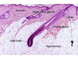
Wet Mount PT: 2006A Micrograph 1. Evaluate images 1-a, 1-b, and 1-c together as if different fields from the same patient Identify cellular images identified as: 1 2 3 Enter results on result sheet. Micrograph 1-a. 3. 1. 2. Micrograph 1-b. 1. 3. 2. Micrograph 1-c. 1. 3.

E N D
Wet Mount PT: 2006AMicrograph 1 • Evaluate images 1-a, 1-b, and 1-c together as if different fields from the same patient • Identify cellular images identified as: • 1 • 2 • 3 • Enter results on result sheet
Micrograph 1-a 3 1 2
Micrograph 1-b 1 3 2
Micrograph 1-c 1 3
Wet Mount PT: 2006A Micrograph 2 • Evaluate images 2-a, 2-b, and 2-c together as if different fields from the same patient • Identify cellular images identified as: • 1 • 2 • 3 • Enter results on result sheet
Micrograph 2-a 1 2
2 Micrograph 2-b 3 2
Micrograph 2-c 2 3
Wet Mount PT: 2006A Micrograph 3 • Evaluate images 3-a, 3-b, 3-c, and 3-d together as if different fields from the same patient • Identify cellular images identified as: • 1 • 2 • 3 • Enter results on result sheet
Micrograph 3-a 1 2
Micrograph 3-c Low Power 3 3 3
High Power Micrograph 3-d 3 3
Wet Mount PT: 2006A Educational Challenge • Evaluate the following micrographs • These will not be graded!
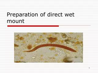
Pseudohyphae vs. Artifact • Fibers are generally larger in size that pseudohyphae • Pseudohyphae have parallel sides with a consistent dimension between the sides while fibers show variable widths along the fiber. • Fibers tend to be birefringent; i.e, they change color when focusing up and down on the object. Colors are often gold or blue and result from the microscope light being refracted by the fiber.
Sign & Date Result Sheet • Testing Person • Site Coordinator • Send results to lab director within 10 days of electronic notification of PT challenge