Download
1 / 1
10 likes | 155 Views
New Multi-channel Measurement Near IR Light For Optical Brain Imaging Jean-Claude Vouakouanitou , Cognitive Neuroscience Lab. Encino, CA 91436. Conclusions. Introduction. Method. Implementation of Beer’s Law. Near Infrared Spectroscopy (NIRS)

E N D
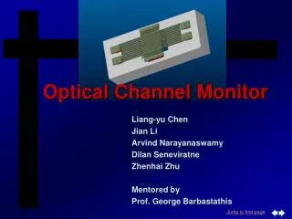
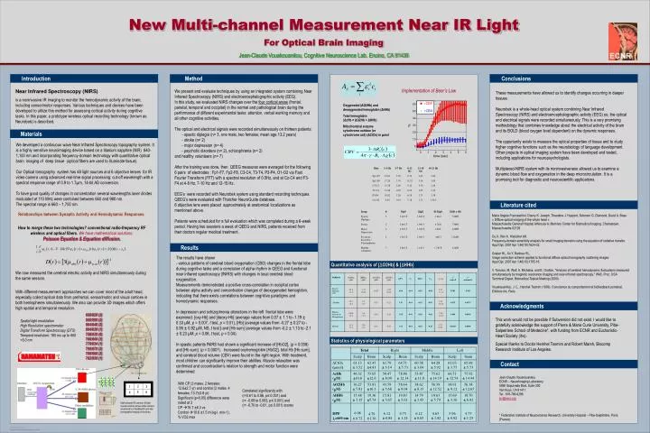
New Multi-channel Measurement Near IR Light For Optical Brain Imaging Jean-Claude Vouakouanitou, Cognitive Neuroscience Lab. Encino, CA 91436 Conclusions Introduction Method Implementation of Beer’s Law Near Infrared Spectroscopy (NIRS) is a noninvasive IR imaging to monitor the hemodynamic activity of the brain, including sensorimotor responses. Various techniques and devices have been developed to utilize this method for assessing cortical activity during cognitive tasks. In this paper, a prototype wireless optical recording technology (known as Neurobek) is described. We present and evaluate techniquesby using an integrated system combining Near Infrared Spectroscopy (NIRS) and electroencephalographic activity (EEG). In this study, we evaluated NIRS changes over the four cortical areas (frontal, parietal, temporal and occipital) in the normal and pathological brain during the performance of different experimental tasks: attention, verbal working memory and all other cognitive activities. The optical and electrical signals were recorded simultaneously on thirteen patients: - spastic diplegia (n= 3, one male, two females; mean age 13.2 years) - stroke (n= 2) - major depression (n= 4) - psychotic disorders (n= 2), schizophrenia (n= 2) and healthy volunteers (n= 7) After the training was done, then QEEG measures were averaged for the following 6 pairs of electrodes : Fp1-F7, Fp2-F8, C3-C4, T3-T4, P3-P4, O1-O2 via Fast Fourier Transform (FFT) with a spectral resolution of 0.5Hz, and at Cz-C4 and F3-F4 at 4-8 Hz, 7-10 Hz and 12-15 Hz. EEG’s were recorded with Neurobek system using standard recording techniques. QEEG’s were evaluated with Thatcher NeuroGuide database. 6 objective lens were placed approximately at anatomical localizations as mentioned above. Patients were scheduled for a full evaluation which was completed during a 6-week period. Having two sessions a week of QEEG and NIRS, patients received from their doctors regular medical treatment. These measurements have allowed us to identify changes occurring in deeper tissues. Neurobek is a whole-head optical system combining Near Infrared Spectroscopy (NIRS) and electroencephalographic activity (EEG) so, the optical and electrical signals were recorded simultaneously. This is a very promising methodology that combines knowledge about the electrical activity of the brain and its BOLD (blood oxygen level dependent) on the dynamic responses. The opportunity exists to measure the optical properties of tissue and to study higher cognitive functions such as the neurobiology of language development. Other projects in optical imaging system have been developed and tested, including applications for neuropsychologists. Multiplexed-NIRS system with its noninvasiveness allowed us to examine a dynamic blood flow and oxygenation in the deep microcirculation. It is a promising tool for diagnostic and neuroscientific applications. Oxygenated (ΔO2Hb) and deoxygenated hemoglobin (ΔHHb) Total hemoglobin (ΔcHb = ΔO2Hb + ΔHHb) Mitochondrial enzyme cytochrome oxidase (or cytochrome aa3) (ΔCtOx) in µmol Materials We developed a continuous wave Near Infrared Spectroscopy topography system. It is a highly sensitive neuroimaging device based on a titanium sapphire (NIR) 840-1,100 nm and incorporating frequency-domain technology with quantitative optical brain imaging of deep tissue (optical fibers are used to illuminate tissue). Our Optical tomography system has 48 light sources and 6 objective lenses for IR video camera using advanced real-time signal processing: cut-off wavelength with a spectral response range of 0.9 to 1.7µm, 14-bit AD conversion. To have good quality of changes in concentration several wavelengths laser diodes modulated at 110 MHz were combined between 660 and 980 nm. The spectral range is 660 – 1,700 nm. Literature cited Relationships between Synaptic Activity and Hemodynamic Responses. How to merge these two technologies?conventional radio-frequency RF wireless and optical fibers.We have mathematical solutions: Poisson Equation & Equation diffusion. We now measured the cerebral electric activity and NIRS simultaneously during the same session. With different-measurement approaches we can cover most of the adult head, especially collect optical data from prefrontal, sensorimotor and visual cortices in both hemispheres simultaneously. We also can provide 3D images which offers high spatial and temporal resolution. Maria Angela Franceschini, Danny K. Joseph, Theodore J. Huppert, Solomon G. Diamond, David A. Boas « Diffuse optical imaging of the whole head » Massachusetts General Hospital Athinoula A. Martinos Center for Biomedical Imaging, Charlestown, Massachusetts 02129 Gu X. Ren K. Hieischer AH. Frequency-domain sensitivity analysis for small imaging domains using the equation of radiative transfer. Appl Opt. 2007 Apr 1;46(10):1624-32. Graber HL, Xu Y, Barbour RL. Image correction scheme applied to functional diffuse optical tomography scattering images. Appl Opt. 2007 Apr 1;46(10):1705-16 V. Toronov, M. Wolf, A. Michalos, and E. Gratton, "Analysis of cerebral hemodynamic fluctuations measured simultaneously by magnetic resonance imaging and near-infrared spectroscopy," WA5, Proc. OSA Technical Digest, Biomedical Topical Meeting (2000) Vouakouanitou, J. C., Hershel Toomim (1999). Conscience du comportement et biofeedback pondéral, Editions ihs, Paris. Results The results have shown - various patterns of cerebral blood oxygenation (CBO) changes in the frontal lobe during cognitive tasks and a correlation of alpha rhythm in QEEG and functional near infrared spectroscopy (fNIRS) with changes in local cerebral blood oxygenation. Measurements demonstrated a positive cross-correlation in occipital cortex between alpha activity and concentration changes of deoxygenated hemoglobin, indicating that there exists correlations between cognitive paradigms and hemodynamic responses. In depression and schizophrenia alterations in the left frontal lobe were examined: [oxy-Hb] and [deoxy-Hb] (average values from 0.07 ± 1.1 to -1.19 ± 0.33 µM, p = 0.007, t test, p = 0.01), [Hb] (average values from -0.27 ± 0.27 to -0.95 ± 0.92 µM, NS, t test ) and [Hb sum] (average values from -0.2 ± 1.13 to -2.1 ± 0.23 µM, p = 0.06, t test, p = 0.04). In spastic patients fNIRS had shown a significant increase of [HbO2], (p = 0.008) and [Hb sum], (p = 0.0001). Increased oxyhemoglobin [HbO2], total Hb [Hb sum], and cerebral blood volume (CBV) were found in the right region. With treatment, most children can significantly improve their abilities. Muscle relaxation was confirmed and cocontraction’s relation to strength and motor function were determined. Quantitative analysis of [ΔO2Hb] & [ΔHHb] Acknowledgments 660/820 (2)660/980 (2)664/848 (2)664/830 (1)758/830 (8)760/840 (3)779/834 (1)780/870 (2)780/960 (2)782/830 (1) This work would not be possible if Subversion did not exist. I would like to gratefully acknowledge the support of Pierre & Marie Curie University, Pitie-Salpetriere School of Medecine*, with funding from ECNR and Eucharistic-Heart Society (ihs). Special thanks to Doctor Hershel Toomim and Robert Marsh, Biocomp Research Institute of Los Angeles. Spatial light modulation High Resolution spectrometer Digital Transform Spectroscopy (DTS) Temporal resolution: 160 ms up to 660 <5.0 nm Statistics of physiological parameters Contact Jean-Claude Vouakouanitou ECNR – NeuroImaging Laboratory 5990 Sepulveda Blvd, Suite 200 Van Nuys, CA 91411 Tel.: 818-786-6286 jcv@ecnr.org * Federative Institute of Neuroscience Research, University Hospital – Pitie-Salpêtrière, Paris (France). With CP (3 males, 2 females; 12.6±2.7 yr) and control (3 males, 4 females; 13.7±2.8 yr) Significant (p<0.05) difference were noted at 3 CP 15.7 ±6.3 vs Control 10.6 ±1.5 ml.kg-l. min-1), % VO2 max 2 3 1 Correlated significantly with (r=0.61 to 0.86, p≤ 0.001) and (r= -0.69 to 0.063, p≤ 0.001) and (r= -0.70 to -0.61, p≤ 0.001) scores 5 6 4 Multi-channel IR camera: 48 light sources and six lenses video camera are placed in a headband to provide tomographic imaging of the brain.