Download
1 / 50
950 likes | 5.45k Views
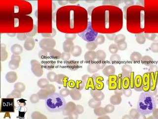
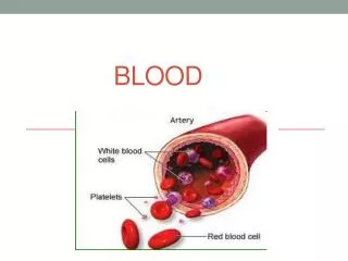
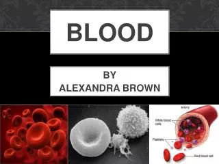
I. Composition & Function of Blood. A. Components 1. Plasma - Connective tissue in which formed elements are suspended in a fluid matrix called plasma Formed elements: 1. Erythrocytes: red blood cell 2. Leukocytes: white blood cells

E N D
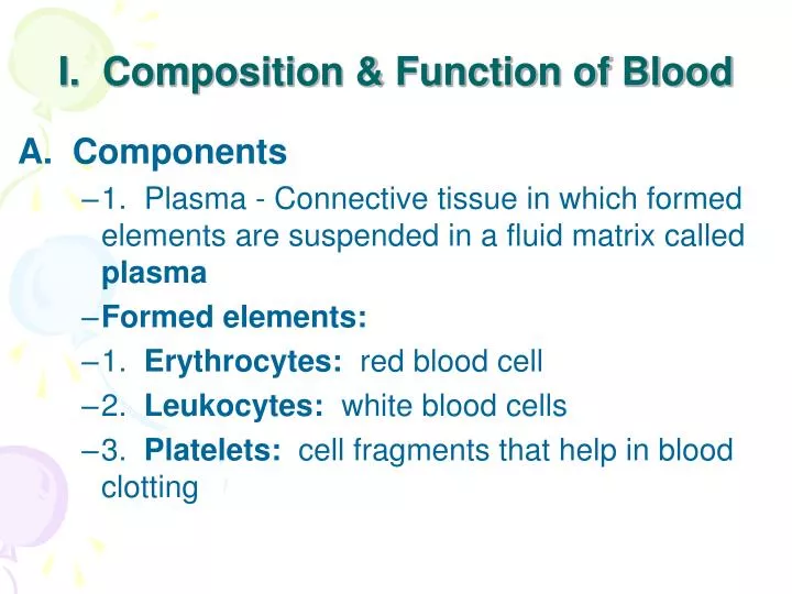
I. Composition & Function of Blood • A. Components • 1. Plasma - Connective tissue in which formed elements are suspended in a fluid matrix called plasma • Formed elements: • 1. Erythrocytes: red blood cell • 2. Leukocytes: white blood cells • 3. Platelets: cell fragments that help in blood clotting
B. Physical Characteristics: • 1. Blood is slightly alkaline; pH 7.35 to 7.45 • 2. Hematocrit: % of RBC's; normal is 45%
C. Functions • Transport • a. Transports hemoglobin that carries oxygen from the lungs to the tissues • b. Contains carbonic anhydrase that catalyzes the reversible reaction between carbon dioxide and water. So the water of the blood transports CO2 from tissues to the lungs (HCO3-) • c. Hormones to target tissues • Regulation • a. Body temperature by absorbing and distributing heat • b. Maintaining normal pH in body tissues • c. Maintains fluid volume in circulatory system; NaCl, salts, blood proteins (albumin) • Protection • a. Clot formation to prevent blood loss • b. Preventing infection (antibodies, WBC's)
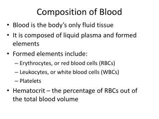
II. Blood Plasma • A. General 1. 90% water 2. 100+ dissolved solutes including nutrients, gases, hormones, wastes, ions, proteins 3. Plasma proteins: a. Albumins: 60%; carrier molecule, buffer, plasma osmotic pressure with Na+ b. Fibrinogen: clotting protein c. Globulins: antibodies Function: blood clotting, defense, hormones, enzymes, carriers for different substances.
1. Name the 3 types of blood cells. • 2. Which cells are complete cells (have a nucleus)?
III. Three Cellular Elements • B. Erythrocytes - red blood cells (RBC) • Leukocytes - white blood cells (WBC) • Platelets - cell fragments • Only leukocytes are complete cells • erythrocytes have no nuclei and platelets are cell fragments 2. Most survive in the bloodstream for only a few days 3. Most do not divide but are renewed in the bone marrow
C. Erythrocytes 1. General • a. Biconcave shape • b. Lack a nucleus • c. Job is transport of oxygen from the lungs to the cells and carbon dioxide from cells to the lungs; Hemoglobin: fills 97% of cytoplasm for gas transport • d. Shape: flexible, stackable, increased surface area for gas exchange • e. Lack mitochondria but do have cytoplasmic enzymes that produce ATP
2. Hemoglobin • a. 4 globin (protein) chains centered around a heme group • b. 250 million Hb molecules per RBC & each can carry four oxygen molecules so each RBC can carry 1 billion molecules of O2 • c. Containment in RBC's prevents breaking into fragments & contributes to blood viscosity & osmotic pressure • d. Normal values for hemoglobin are 13-18g/100ml in adult males, and 12 to 16g/100ml in adult females
3. Erythropoiesis (RBC formation) • Begins in early embryonic development (in yolk sac) and continues throughout life. • b. Red cell-producing marrow cells are called hematopoietic stem cells (hemato=blood; poiesis=to make): • c. Blood cells (except lymphocytes) are descendants of a single precursor cell Stimulated by Erythropoietin
Bone Marrow 1 oz. of new blood with 100 billion new cells per day Red cell-producing marrow cells are hematopoietic stem cells In adults, only the spine, ribs, pelvis, bodies of vertebrae, portions of skull, and proximal ends of long bones produce blood cells.
4. Regulation & Requirements • 1) Hormonal control of erythrocyte production • a) Erythropoietin - a glycoprotein secreted by kidneys when oxygen levels are decreased • Stimulates red marrow “RBC’s” to mature • 2.5 million erythrocytes/second 2) Requirements • a) Iron: 65% of body’s supply is in hemoglobin • b) Vitamins: B12 & folic acid for DNA synthesis • 3) Fate & Destruction • a) Erythrocytes cannot synthesize proteins, grow, or divide. Circulate about 120 days before being destroyed. • b) Self-destruct in the red pulp of spleen and liver
4. Disorders • a. Anemias: deficiency of hemoglobin, too few red blood cells, (hemoglobin) so decreased oxygen-carrying capacity: • 1) Low number of RBCs • a) Hemorrhagic anemia • b) Aplastic anemia - bone marrow failure • 2) Low Hemoglobin content • a) Iron-deficiency anemia; excretion exceeds intake • b) Athlete’s anemia; vigorous exercise can expand blood volume. • c) Pernicious anemia; deficiency of vitamin B12 • 3) Abnormal Hemoglobin: genetic • a) Sickle-cell anemia; change in one of 287 amino a. • b. Polycythemias: high RBC counts; blood viscosity increases causing it to flow sluggishly; often in bone marrow cancer
4. Blood doping • In the late 1980s erythropoietin, a growth factor that stimulates the formation of RBCs, became popular. • Recombinant DNA technology made it possible to produce EPO economically; approved in US and Europe as a pharmaceutical product for the treatment of anemia resulting from renal failure or cancer chemotherapy.
Leukocytes - white blood cells: Formed in lymphoid tissue(lymph nodes, tonsils, spleen, thymus) and then transported in blood to different parts of the body. Attracted to certain chemicals released by damaged cells or other leukocytes and move by amoeboid motion Provide a rapid and potent defense against infection Are the only formed elements of blood that are complete cells
Leukocytes - white blood cells 5 types of mature white blood cells (leukocytes): lymphocytes, monocytes, neutrophils, eosinophils, basophils.
Questions • Jack has appendicitis. What cell types may be increased in his blood? • Kay has a viral infection. What cell types may be increased in her blood?
Answers • Jack has appendicitis. What cell type may be increased in his blood? Neutrophil • Kay has a viral infection. What cell type may be increased in his blood? Lymphocyte
C. Leukocytes: • a. Less than 1% of blood volume; 4,000-11,000 WBCs per cubic millimeter of blood • b. Leukocytosis: WBC count over 11,000; normal response to invasions as the body speeds up production • c. Diapedesis: unlike red blood cells that are confined to the bloodstream, WBCs can leave capillaries and enter loose connective and lymphoid tissues • d. Grouped • Granulocytes - contain cytoplasmic granules • Agranulocytes - lack granules
2. Granulocytes • Neutrophils • Most numerous : > 1/2 of WBC population or 50-70%. • a. A lilac stained cytoplasm and a lobed nucleus (polymorphonuclear) • b. Chemically attracted to sites of inflammation • c. Very effective phagocytes of bacteria and viruses which they attack and destroy even in circulating blood • d. Are the bodies bacteria slayers and numbers increase during bacterial infections
b. Eosinophils • 2-4% of blood leukocytes • Blue-red nucleus with two lobes • Attack parasitic worms, attach by surface molecules and release substances from their cytoplasm to the parasite’s surface • 4) Also involved in allergic response by inactivating inflammatory chemicals
c. Basophils • 1) Rarest white blood cell; 0.5-1% of leukocyte population • 2) Cytoplasm releases histamine, an inflammatory chemical that vasodilates blood vessels and attracts other white blood cells to the inflamed site. (Antihistamines counter this effect)
3. Agranulocytes • a. Lymphocytes • 1) Second most numerous • 2) Nucleus fills cytoplasm • 3) Small percentage in blood, most in lymphoid tissues • 4) Two major types: • T cells - control immune responses • B cells - produce antibodies • b. Monocytes • 1) Largest (18 µm); U-shaped purple stained nucleus • 2) Differentiate into macrophages in tissues; defend against viral and bacterial parasites
4. Production & Lifespan of Leukocytes • Leukopoiesis: • Hormonally stimulated by cytokines - proteins that control the immune response. • Cytokines are released by active white blood cells and stimulate the production of additional neutrophils and macrophages. • 5. Leukocyte Disorders • a. Leukemias: group of diseases of abnormal growth and development of white blood cells
Renegade leukocytes that remain unspecialized and divide out of control, impairing normal bone-marrow function Leukemia
D. Platelets a. Function: contain chemicals that act in blood clotting b. Cell fragments split off a parent cell - megakaryocytes. Thrombopoietin: a glycoprotein that regulates growth and maturation of megakaryocytes
List and explain the stages of HEMOSTASIS • 1. FORM 3 GROUPS • 2. EACH GROUP ACT OUT EACH STAGE
IV. Hemostasis • Process of keeping blood within the vessels by repairing breaks. • A. Involves three phases: A cascade of events: • 1. Vascular spasm • 2. Platelet plug formation • 3. Coagulation - blood clotting • B. Vascular spasm • 1. Vasoconstriction caused by injury to smooth muscle of vessel walls. WHY? A constricted artery reduces blood loss.
C. Platelet Plug Formation • 1. Endothelium damage exposes collagen fibers • 2. Platelets swell & form spiked processes and become sticky & adhere to exposed collagen • 3. Then the platelets release serotonin that enhances vascular spasm to attract more platelets • 4. Positive feedback • 5. Platelet plug is formed (Aspirin blocks this process)
1. Blood is transformed from liquid to a gel 2. Prothrombin converts a plasma protein called prothrombin > thrombin, an enzyme 3. Thrombin catalyzes fibrinogen present in the plasma to a fibrin mesh that traps blood cells and seals the hole until the vessel can be repaired 4. Clot formation complete in 3-6 minutes CLOTTING
Clot Retraction & Repair: • 1. Within 30 minutes, the clot is stabilized • 2. Actomyosin: contractile protein complex in platelets that contracts and pulls on fibrin strands, squeezing serum from mass and drawing the edges or the vessels together • 3. Platelet-derived growth factor (PDGF) stimulates smooth muscle & fibroblasts to divide & rebuild wall
Fibrinolysis - removal unneeded clot when healing has occurred. Vessels would become blocked without this process. • 1. Plasmin: a fibrin-digesting enzyme is produced to dissolve fibrin. • 2. Begins within two days and continues over several days
H. Disorders of Hemostasis • a. Thrombus: clot in an intact vessel • b. Embolus (wedge): free floating clot • c. Arteriosclerosis, inflammation may roughen endothelium
I. Clinically Prevent Clotting • d. Aspirin - blocks platelet aggregation • e. Coumadin - interferes with vitamin K • 2. Bleeding Disorders • a. Platelet deficiency • b. Impaired liver function • c. Hemophilias
V. Transfusion & Blood Replacement • A. Human Blood Groups - ABO and Rh 1. RBCs have antigens (agglutinogens) on surface that identify 2. RBC antigens promote agglutination so are called agglutinogens. • One person’s RBC protein may be recognized as foreign if transfused into someone else and will agglutinate or clump. • Produce antibodies against antigens not present on cell
C. Rh Blood Groups • 1. Eight antigens (agglutinogens) called Rh factors; 3 common • C, D, E • 2. 85% Americans are Rh+ • RBCs carry the Rh antigen • 3. Rh antibodies only formed in blood upon exposure (not preformed like ABO) • 4. Hemolysis occurs after the second exposure and thereafter
Example If the baby's Rh positive blood enters a mother who is Rh Negative, then her immune system sees the cells as 'incompatible' or 'foreign' and will subsequently produce anti-rhesus antibodies to try to destroy them for her own self-protection. If she has another pregnancy where the baby is Rh positive, then these antibodies that have formed will pass into the baby's bloodstream via the placenta and attack the red blood cells of the baby. Once these naturally created anti-rhesus antibodies are present in the mother's bloodstream, they will remain for life. Give Rho Gam that masks the agglutinin component so it does not recognize its presence and activation will not occur.
B. Common Lab Evaluations • 1. Total white cell count: total number of all types of white blood cells • 2. Differential and absolute WBC Count: estimates the relative numbers of the 5 types of white cells • 3. Hematocrit :% of total blood volume that is packed red blood cells • 4. Platelet count: ability to clot • 5. Blood chemistry: ammonia (liver & renal), amylase (pancreas), aspartate (cell damage due to MI, liver disease, drugs), bilirubin (liver), blood urea nitrogen (kidney), blood gases, Igs, lipoprotein, lactic acid, protein, potassium, sodium, triglycerides, uric acid