Download
1 / 15
230 likes | 1.08k Views
Comparison of Microwave and Radiofrequency Ablation of Hepatic Tissue in a Porcine in vivo Model. Andrew Wright MD, Chris Johnson, Fred Lee Jr. MD, and David Mahvi MD. Departments of Radiology and Surgery University of Wisconsin Hospital. Microwave Ablation.

E N D
Comparison of Microwave and Radiofrequency Ablation of Hepatic Tissue in a Porcine in vivo Model. Andrew Wright MD, Chris Johnson, Fred Lee Jr. MD, and David Mahvi MD Departments of Radiology and Surgery University of Wisconsin Hospital
Microwave Ablation • Theoretical advantages over radiofrequency ablation • No ground pad • Not limited by tissue charring and impedence changes • Larger zone of active heating • Use of Multiple Probes
Hypothesis • Because microwave and radiofrequency ablation are both heat based, there will be no difference in ablation size or lesion pathology between the two technologies
Methods • Microwave Ablation • Vivant Medical prototype system • 10 minute ablation, 40 Watts • Radiofrequency Ablation • RITA Medical Systems Starburst • 10 minute ablation, 3cm deployment 100oC target temperature
Microwave Ablation System • Vivant Medical • 13g, 15cm dipole antenna • 915MHz generator • Fiberoptic temperature monitor
Radiofrequency Ablation System • RITA Medical • 14g, 15cm expandable array • 460 kHz generator • Integrated thermocouple
Treatment Protocol • Nineteen crossbred female swine • 0, 2, and 28 day survival groups • Mix of MW and RF lesions in each animal • Pathology, serial CT scanning • 28 Day survival group • 4 MW or 4 RF lesions in each animal • Pathology, serial lab draws
Lesion Volume * * * p=.02
Lesion Length * ▪ ▪ ◦ * ◦ * p<.001 ▪ p=.02 ◦ p<.001
Laboratory Data • No significant difference in AST, ALT, LDH, Alkaline Phosphatase, WBC, or HCT * * p<0.001
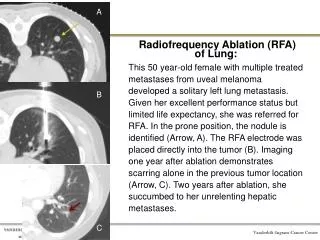
48o Pathology RFA MW Immediate 4 weeks
CT Imaging 48 Hours 4 Weeks
Summary • MW lesions longer than RF • Pathological and radiologic characteristics similar between RF and MW ablation • Both MW and RF cause thrombocytopenia at 48 hours • MW ablation technically easier than multiple-prong RF ablation
Conclusions • MW creates similar zones of coagulation necrosis as RF • MW ablation has several theoretical advantages over RF • Larger zone of active heating • Ability to use multiple probes