Download
1 / 15
150 likes | 277 Views
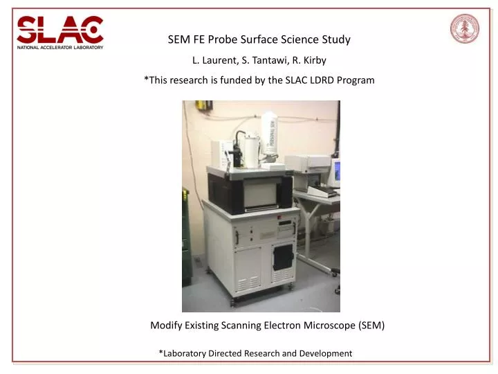
SEM FE Probe Surface Science Study L. Laurent, S. Tantawi, R. Kirby *This research is funded by the SLAC LDRD Program. Modify Existing Scanning Electron Microscope (SEM). *Laboratory Directed Research and Development. AFM Field Emission Probe SLAC-TN-03-056 (2003).

E N D
SEM FE Probe Surface Science Study L. Laurent, S. Tantawi, R. Kirby *This research is funded by the SLAC LDRD Program Modify Existing Scanning Electron Microscope (SEM) *Laboratory Directed Research and Development
AFM Field Emission Probe • SLAC-TN-03-056 (2003) Measurement Problems and Restrictions: • Small Samples (~size of a quarter) • Very Small Diagnostic Area (mm’s) • Non Vacuum Chamber • Tip and Sample Oxidation • Tip and Surface Contamination • Unknown Gap Distance Robert E. Kirby / Lisa Laurent
Field emission probe will detect features that exhibit premature field emission. The surface imaging capability of the SEM and elemental x-ray analysis can then be used to obtain topographical and material information about these emission sites. SLAC SEM has one of the largest commercially available vacuum chambers which allows us to analyze cathodes, windows, and some high gradient structures. The FE probe assembly has three degrees of freedom, and nanometer resolution. SEM has XYZ stage, tilt, and angular rotation. This wide latitude of sample and probe placement will allow field emission measurements with distance and angle as variables.
A Brief History of Field Emission, Emitters, and Imaging In 1897, R.W. Wood discovered field emission while conducting dc vacuum experiments. In 1928, Fowler and Nordheim derived an equation describing the field emission current density in relationship to electric field strength. In 1936, Ahearn proposed that field emission currents consisted of pencil-like streams of electrons emitted from a few minute areas, in contrast to being emitted uniformly over the entire surface. In 1963, Little and Whitney experimentally verified that field emission originates from multiple sites using a phosphor-coated anode.
High Voltage Vacuum Insulation: Basic Concepts and Technological Practice (Edited by Rod Latham) Transparent Anode. Plane-parallel Electrodes. Discrete localized emission sites Gap field gradually increased.- Arc location at pre-breakdown emission site Post breakdown event frequently resulted in the creation of many additional new emission sites. (1995)
SEM Field Emission Experiments In 1975, B.M. Cox used a stainless steel planar anode with a “probe hole” situated in a scanning electron microscope. An electrode behind the probe hole collected the current. Poor resolution (~10mm’s) – not able to identify topographical features. In 1981, Athwal and Latham used a pointed anode probe SEM with limited elemental analysis capabilities. Lacked stage precision to locate FE sites. In 1994, M. Jimenez, R. Noer used flat tip (diam.=80mm) truncated cone installed in SEM. Limited elemental analysis capabilities. Analysis focused on primarily niobium cathodes. Superficially added particles to surface.
Cathode Research The performance of cathodes is highly dependent by the physics and chemistry taking place on the surface. The FE SEM system will be used to identify possible causes of non-uniform cathode emission. SSRL Dispenser Cathode
Cathode Research The current probe will provide diagnostic support to help understand emission physics and in advancing next generation photocathode high brightness beams. We will use the current probe system to find features that may be responsible for breakdown. Laser Cleaning LCLS Photocathode Inside SLAC SEM vacuum Chamber Breakdown
High Gradient Research -- Clamped vs Brazed -- Identify the field emission characteristics of structures before and after they have been tested to help understand the relationship between surface physics and rf breakdown. Clamped Single Cell Structure Brazed Single Cell Structure Clamped HDX11
W Iris Insert - CERN HIP Cu - KEK High Gradient Research -- Materials -- TiN Coated Cu - KEK CuZr-SLAC CuZr-SLAC Moly Plated Cu - Frascatti CuCr CuZr-CERN
High Gradient Research -- Surface Finish -- Etch Pits Pre-RF Electro-polished Post-RF
LDRD FE Probe SEM Program Overview and Current Status • Design Stage: • Assembly drawings are completed. Field emission probe assembly and auxillary • components have been specified. • Ordering Parts: • A capital equipment purchase order is in process and we expect to receive shipment • of all components by 03/15/11. • Installation • While awaiting delivery we will modify the SEM sample stage box to include • mounting holes and a flange opening for the FE holder and cable flange. • Initial Testing and Calibration of the System on Planar Test Samples (e.g. Copper Coupons): • Begin test sample measurements and calibration by 5/1/11.
LDRD FE SEM Program Overview and Current Status–pg 2 • Analyze Existing (already tested) Cross Sectioned Accelerator Structures and Cathodes. • Evaluate working with non planar geometries. • Surface Science Study to Include Iterative Process (before and after rf testing and/or beam operation): • High Gradient Program: Clamped structures • Cathode Research: Demountable Cathodes
New Higher Resolution SLAC SEM in 2012…?? SLAC SEM is reaching end of life status. The goal is to optimize the system in SLAC’s current SEM and than move the system to a new SEM in the future when funding is available. New SEM was approved in 2010. Purchase requisition was halted days before order was placed due to the budget being on continuing resolution. Present day SEM’s have significantly better capabilities and resolution than SLAC’s current SEM.
Summary • In high gradient structures and cathode development we are faced with challenging physics that will likely only be overcome by increasing our surface science capabilities. • The current probe coupled with SEM imaging and EDX spectrometry will provide additional diagnostic support to help understand surface and emission physics. • The SEM field emission probe will be available for a wide variety of applications. • We hope to increase the field emission probe system capabilities by installing it into a new higher resolution SEM as soon as funding is available.