Download
1 / 70
710 likes | 933 Views
Immune Cells Paul Zhou Institute Pasteur of Shanghai, CAS. Overview of lymphatic system.

E N D
Lymphatic system1. Consist of organs, nodes and vessels2. Distribution throughout of body 3. Interface with environment4. Circulating throughout body (uni-directional, regional, connected to blood circulation system)5. Immune privilege sites
Organ and tissues in immune system1. primary lymphoid organs: bone marrow and thymus- ontogeny and development of lymphocytes and some non-lymphocyte cells (acquisition of capability of antigen recognition, becoming effector/memory cells, and homing)2. secondary lymphoid organs: spleen, lymph nodes, tonsils and Peyer’s patches- antigen encounter and immune activation (and effector response)3. tertiary lymphoid tissues: skin and mucosa- sites of antigen acquisition and processing and effector response
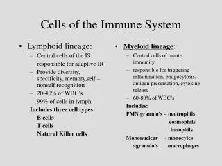
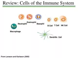
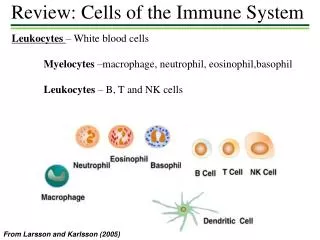
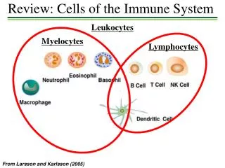
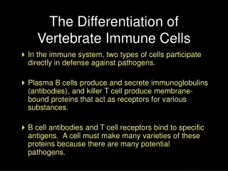
Cells in immune system1. Lymphoctyes: T, B and NK cells- determine the immune specificity and orchestrate the effector response (T and B cells); both innate and adaptive immunity (NK cells)2. Non-lymphocytes: monocytes/macrophages, dendritic cells and neutrophils, basophils, eosinophils and mast cells- interact with lymphocytes and play a critical role in the antigen presentation and the mediation of immunologic functions 3. Specialized epithelial and stromal cells- provide the anatomic environment in which immunity occurs
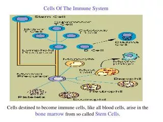
Important features in immune cells1. Developed from hematopoietic stem cells- ontogeny and lineage differentiation2. Replenished throughout life- proliferation, differentiation and maturation, circulation and migration3. Have great potential in response to various insults in antigen specific or non-specific manner- activation, proliferation, maturation, circulation and migration4. Activate locally and then migrate (or circulate)5. Expand and contract during immune response6. Cross talk among immune cells and between immune and non-immune cells
Leukocytes in blood Cell number/μl Range _______________________________________ Whole blood cells 7,400 4,500 - 11,000 (leukocytes) Neutrophils 4,000 1,800 - 7,000 Eosinophils 200 0 - 450 Basophils 40 0 - 200 Lymphocytes 2,500 1,000 - 4,800 Monocytes 300 0 - 800
Topics to be covered in this lecture • Primary lymphoid organs: ontogeny of immune cells • Secondary lymphoid organs: sites of immune activation • Tertiary lymphoid tissues: the sites of antigen acquisition and effector immune response • Migration and homing of immune cells
Bone marrow1. Cellular content of bone cavity2. A reservoir for HSCs3. A site of B cell lymphopoiesis throughout life4. sIgM expressing B cells generated in bone marrow migrate to spleen for further maturation
B-2 cells B-1 cells_________________________________________________Adult vs. fetal adult fetalPercentages 95 5Surface markers CD19/CD45RA CD19/CD45RA CD11b+sLgMhighsLgDlowIg repertoire highly diverse limited diverseAntigens T dependent T independent protein in nature variety including carbohydrateRequirement of Th cells yes noIg class switching yes noAffinity maturation yes no
Trafficking of thymocytes in T cell development and selection
Lymphocyte distribution (% of total) __________________________________ Blood Lymph nodes Spleen B cells 10 - 15 20 - 25 40 - 45 Th cells 50 - 60 50 - 60 40 - 50 Tc cells 20 - 25 15 - 20 10 - 15 NK cells about 10 rare about 10
Histology of lymph node1. capsule2. HEV3. cortex – para-cortex (T zone) and 4. germinal center 5. sinus6. in and out lymph node
T cell activation and cellular immune response • Two major subsets: α/β TCR and γ/δ TCR • Two lineages in α/β TCR T cells: CD4 and CD8 - differ in antigen recognition as well as regulatory and effector functions • Two subtypes in CD4 T cells: Th1 and Th2 - differ in cytokine secretion and helper functions • Naïve, effector (activated), and memory T cells • Recognize processed antigen/MHC complex on the surface of APCs • T cells become activated through signals 1 and 2 • Activated (effector) T cells become proliferative through IL-2 and IL-2R autocrine and paracrine loop and migrated to infection site(s) • Activated T cell contraction • Central memory and effector memory T cells
T Cell Receptors • TCR consists of two subsets: α/β and γ/δ. • Through somatic gene arrangement, the TCR displays extreme sequence diversity. • TCR expresses on the surface of T cells in a clonal fashion, in which to a given T cells only one pair of α/β or γ/δ TCR is expressed. • α/β or γ/δ TCR itself does not mediate signaling. Instead it is intimately associated with CD3 complex (γδ2ε2ζ). It is the latter that mediates TCR signaling. • The signal mediated through TCR/CD3 complex is called the signal one.
Co-stimulatory molecules between T cells and antigen presenting cells
Effector functions of T cells • Early changes • Changes in pH • Changes in membrane potential (MTT assay) • Fluxes in cyclic nucleotides and calcium (Ca influx assay) • Late changes • Cyto-skeletal changes (morphologic change) • Activation of the cytolytic mechanism (51Cr and enzyme release assays) • Gene regulation: CD25, CD69, IL-2, IL-3, IFNγ, GM-CSF, CTLA-4, MHC class II, VLA-2, 4F2, transferrin receptors, and insulin receptors, etc. (Elisa, Elispots, intracellular cytokine staining, activation markers) • T cell proliferation (3H-thymidine and CFSE assays as well tetramer staining)
Tertiary lymphoid tissues: the sites of antigen acquisition and effector immune response
Mucosal sites Lymphocyte subsets Distribution (%) Possible functions CD3+ T cells 35–40 CD4+, CD8- 65 Major T-helper cells for mucosal immunity CD4-, CD8+ 30 CTL precursors; regulatory/anergy CD4-, CD8- 2–4 Express gd TCRs Inductive tissues Peyer’s patches Naive 30–40 Circulate within the mucosal system Effector (activated) 30–35 Stimulated through M-cell pathways Memory 30–40 Homing to effector sites B220+ B cells 45–47 Include germinal center where >60% are sIgA+B cells sIgA+ ~8–10 Committed to IgA CD3+ T cells 40–50 CD4+ CD8- ~60–65 Difficult to activate via TCR
Mucosal sites Lymphocyte subsets Distribution (%) Possible functions Lamina propria CD4-, CD8+ ~30–35 Mature CTLs; other subset functions? CD4-, CD8- ~2–5 Express gd TCRs Memory >90 sIgA+ B cells 30–50 IgA plasma cells 10–15 Highest numbers of plasma cells in the mammalian immune system Effector tissues CD3+ T cells 85–95 CD4+, CD8- ~5–8 All express ab TCRs Intraepithelial lymphocytes CD4-, CD8+ ~75–80 2/3 are CD8 aa; 80% gd+; 50% ab+ CD4+, CD8+ ~7–10 All express of ab TCRs CD4-, CD8- ~5–8 All express gd TCRs No B cells/plasma cells
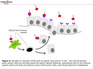
Background information on DCs • Initial discovery of Langerhans cells in skin by Paul Langerhans in 1868 • Dendritic cells (DCs) first described by Steinman and Cohn in mouse spleen in 1973 • Special properties of DCs in initiating immunity (i.e. antigen and pathogen recognition, uptake, process and presentation as well as pathogen dissemination) were discovered after depletion of monocytes, macrophages and B cells • Reside in most peripheral tissues, especially at sites interface with environment; but migrate to draining lymphoid nodes to activate T cells • Can be generated from their precursor cells ex vivo