Download
1 / 1
100 likes | 453 Views
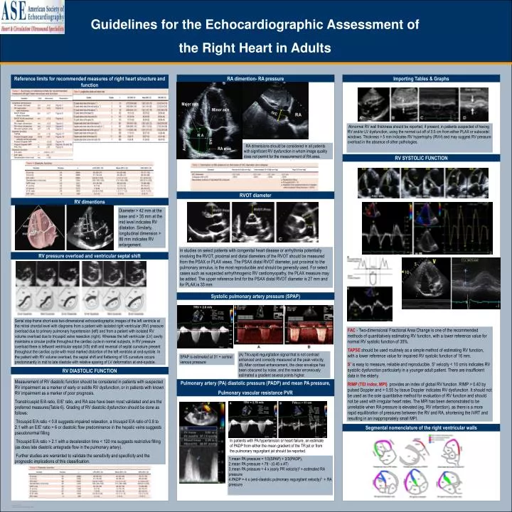
Guidelines for the Echocardiographic Assessment of the Right Heart in Adults. Reference limits for recommended measures of right heart structure and function. RA dimention- RA pressure. Importing Tables & Graphs. Major axis. Minor axis.

E N D
Guidelines for the Echocardiographic Assessment of the Right Heart in Adults Reference limits for recommended measures of right heart structure and function RA dimention- RA pressure Importing Tables & Graphs Major axis Minor axis Abnormal RV wall thickness should be reported, if present, in patients suspected of having RV and/or LV dysfunction, using the normal cut off of 0.5 cm from either PLAX or subcostal windows. Thickness > 5 mm indicates RV hypertrophy (RVH) and may suggest RV pressure overload in the absence of other pathologies. RA area RA dimensions should be considered in all patients with significant RV dysfunction in whom image quality does not permit for the measurement of RA area. RV SYSTOLIC FUNCTION RVOT diameter RV dimentions Diameter > 42 mm at the base and > 35 mm at the mid level indicates RV dilatation. Similarly, longitudinal dimension > 86 mm indicates RV enlargement. In studies on select patients with congenital heart disease or arrhythmia potentially involving the RVOT, proximal and distal diameters of the RVOT should be measured from the PSAX or PLAX views. The PSAX distal RVOT diameter, just proximal to the pulmonary annulus, is the most reproducible and should be generally used. For select cases such as suspected arrhythmogenic RV cardiomyopathy, the PLAX measure may be added. The upper reference limit for the PSAX distal RVOT diameter is 27 mm and for PLAX is 33 mm RV pressure overload and ventricular septal shift Systolic pulmonary artery pressure (SPAP) Serial stop-frame short-axis two-dimensional echocardiographic images of the left ventricle at the mitral chordal level with diagrams from a patient with isolated right ventricular (RV) pressure overload due to primary pulmonary hypertension (left) and from a patient with isolated RV volume overload due to tricuspid valve resection (right). Whereas the left ventricular (LV) cavity maintains a circular profile throughout the cardiac cycle in normal subjects, in RV pressure overload there is leftward ventricular septal (VS) shift and reversal of septal curvature present throughout the cardiac cycle with most marked distortion of the left ventricle at end-systole. In the patient with RV volume overload, the septal shift and flattening of VS curvature occurs predominantly in mid to late diastole with relative sparing of LV deformation at end-systole. FAC- Two-dimensional Fractional Area Change is one of the recommended methods of quantitatively estimating RV function, with a lower reference value for normal RV systolic function of 35%. TAPSE should be used routinely as a simple method of estimating RV function, with a lower reference value for impaired RV systolic function of 16 mm. S' is easy to measure, reliable and reproducible. S' velocity < 10 cm/s indicates RV systolic dysfunction particularly in a younger adult patient. There are insufficient data in the elderly. RIMP (TEI index, MPI) provides an index of global RV function. RIMP > 0.40 by pulsed Doppler and > 0.55 by tissue Doppler indicates RV dysfunction. It should not be used as the sole quantitative method for evaluation of RV function and should not be used with irregular heart rates. The MPI has been demonstrated to be unreliable when RA pressure is elevated (eg, RV infarction), as there is a more rapid equilibration of pressures between the RV and RA, shortening the IVRT and resulting in an inappropriately small MPI. (A) Tricuspid regurgitation signal that is not contrast enhanced and correctly measured at the peak velocity. (B) After contrast enhancement, the clear envelope has been obscured by noise, and the reader erroneously estimated a gradient several points higher. SPAP is estimated at 31 + central venous pressure RV DIASTOLIC FUNCTION Measurement of RV diastolic function should be considered in patients with suspected RV impairment as a marker of early or subtle RV dysfunction, or in patients with known RV impairment as a marker of poor prognosis. Transtricuspid E/A ratio, E/E' ratio, and RA size have been most validated and are the preferred measures(Table 6). Grading of RV diastolic dysfunction should be done as follows: Tricuspid E/A ratio < 0.8 suggests impaired relaxation, a tricuspid E/A ratio of 0.8 to 2.1 with an E/E' ratio > 6 or diastolic flow predominance in the hepatic veins suggests pseudonormal filling Tricuspid E/A ratio > 2.1 with a deceleration time < 120 ms suggests restrictive filling (as does late diastolic antegrade flow in the pulmonary artery). Further studies are warranted to validate the sensitivity and specificity and the prognostic implications of this classification. Pulmonary artery (PA) diastolic pressure (PADP) and mean PA pressure, Pulmonary vascular resistance PVR Segmental nomenclature of the right ventricular walls In patients with PA hypertension or heart failure, an estimate of PADP from either the mean gradient of the TR jet or from the pulmonary regurgitant jet should be reported. 1.mean PA pressure = 1/3(SPAP) + 2/3(PADP), 2.mean PA pressure = 79 - (0.45 x AT) 3.mean PA pressure = 4 x (early PR velocity)² + estimated RA pressure 4.PADP = 4x (end-diastolic pulmonary regurgitant velocity)² + RA pressure