Download
1 / 43
500 likes | 773 Views
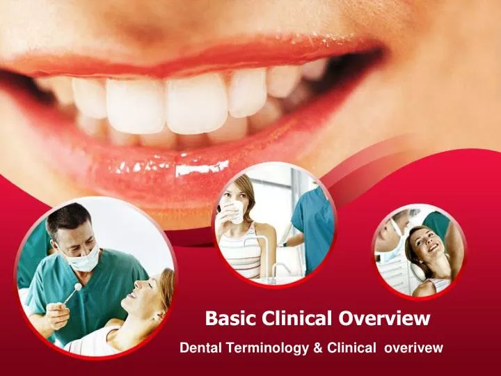
Basic Clinical Overview. Dental Terminology & Clinical overivew. Training. OBJECTIVE: To strengthen your current foundation of dental knowledge in order to improve your confidence assisting patients, both with phone skills and treatment planning. Dental Insurance .

E N D
Basic Clinical Overview Dental Terminology & Clinical overivew
Training OBJECTIVE: To strengthen your current foundation of dental knowledge in order to improve your confidence assisting patients, both with phone skills and treatment planning.
Dental Insurance • There are three categories of dental insurance. They are: • Diagnostic & Preventive • Basic Restorative • Major Restorative The basic rule is 3. Sometimes you will here it referred to has Type A,B,C or Type 1,2,3.
Diagnostic • What procedures are done in the office to help diagnose a problem? • 1. Exam • Emergency/Limited Exam - problem focus • New Patient/Comprehensive Exam - full work up • Periodic Exam - looking for changes since last visit • 2. Dental x-rays • PA’s (Periapicals) - problem focused per tooth • Bitewings - 4 films, one in each quadrant, used to diagnose cavities/fractures, shows molars • Very clear, concise images, lots of detail • Full Mouth Series - 18 single films used to diagnose cavities/fractures, shows all teeth • Very clear, concise images, lots of detail • Panoramic X-ray - wisdom teeth, orthodontics, tumors and cancer, check for erupting teeth • Less detail, not good for cavities!!
X-Rays Continued Single Periapical x-ray to diagnose a problem (As needed)
X-Rays Continued • Bitewing X-rays, (once per year, or every 6 months)
X-Rays Continued • Full Mouth Series (FMX), once in 3 to 5 yrs
X-Rays Continued • Panoramic X-Ray (once in 3 to 5 years)
Preventive Services • What procedures would you consider preventive? (what procedures keep us from getting cavities?)
Preventive Services • Prophy • Routine Dental Cleaning includes scaling up to 3 mm below the gum line and polish
Preventive Services • Sealants • Prevents decay from the occlusal surface of the molars. (chewing surface). Sealants prevent bacteria from collecting in the small grooves. • Insurance companies typically only cover children once every 3 years, or once per lifetime. • Why do sealed teeth still get decay?
Preventive Services • Fluoride Treatment • Strengthens tooth enamel to prevent decay, and can reverse the early stages of cavities. • Reduces tooth sensitivity • Is usually applied as a foam placed in trays, brushed on with a traditional tooth brush, or painted on as a sticky varnish. • Typically insurance will only cover fluoride for children once or twice every 12 months.
Basic Restorative • Usually includes: • Fillings • Endodontics (root canals) • Periodontics (treating the gums) • Oral Surgery (extractions)
Fillings • In order to understand fillings, you must get acquainted with the tooth chart and tooth surfaces.
Tooth Surfaces • There are 5 surfaces on each tooth. When we talk about fillings we call them a 1 - surface, 2-surface, 3 surface or 4+ surface filling. • We use single letters to abbreviate them. For example, an “MOD” would be a 3 - surface filling, because it has 3 letters. • You will be learning the name of each surface accompanied with its location on the tooth. • Occlusal/Incisal • Mesial • Distal • Buccal/Facial • Lingual
Occlusal/Incisal Surface • On the front 6 teeth, we call it the incisal surface, which is used to incise your food. • On the back molars we call it the occlusal surface, which used to chew.
Mesial Surface • The mesial surface is the portion of the tooth closest to the midline, or the middle of your face.
Distal Surface • The distal surface is the portion of the tooth furthest away from the midline, or the middle of your face.
Buccal/Facial • The buccal and facial portion of the tooth is what we could see if you clamped down your teeth, and we removed your cheeks! • When we refer to the front six teeth, we call it the “facial surface.” When referreing to the back molars, it is referred to as the “buccal surface”
Lingual Surface • If you were to clamp down your teeth, the lingual surface is the portion you could lick with your tongue. (the backside of each tooth)
Buccal Occlusal Lingual Incisal Mesial Distal Tooth Surfaces
Time to Practice!! Identify the fillings
Endodontics Why would a tooth require a root canal? Why do teeth with root canals require a crown?
Periodontics • You must first understand gum disease (periodontal disease) before we address the procedures used to treat the condition.
Periodontal Disease • Most people who have periodontal disease do not know it, because it is painless and the gum tissue often covers up most of the problem. Because the gum tissue can disguise signs of periodontal disease, it can be hard for patients to acknowledge they have it. • Bacteria left below the gum line will results in bleeding, bone loss and eventual tooth loss. • This disease is usually caused from lack of flossing, smoking or genetics. • Bone loss cannot be reversed
How is periodontal disease diagnosed? • Your dental hygienist will take routine x-rays to look for bone loss that is hidden under your gum tissue. • She will then take an instrument which will measure the distance from your gum line to the bone. A distance of 3 mm or less indicates healthy bone levels. Anything more indicates loss of bone. • Bleeding during these measurements also indicates active infection, which will lead to continued bone loss. If left untreated - tooth loss is possible. Healthy Bone levels Bone Loss Perio Probe Perio Probing
How is Periodontal Disease Diagnosed? • In addition to dental x-rays and perio probing, your hygienist will also look for other visible signs. • This would include checking for visible red, puffy, swollen or irritated gums and/or gum recession. Gum recession is when the gums pull away from the teeth to avoid plaque and infection. This is much like the automatic response your hand would have after touching a hot stove. Swollen Gums Gum Recession
Treatment of Periodontal disease • In our general dentistry offices, we use a non-surgical approach to treating the disease. This usually entails Scaling and Root Planing, Oral Irrigation with an anti-microbial rinse and Arestin. • After this initial treatment is completed, we recommend patients switch to the Sonicare Tooth Brush, use a strong mouth rinse at home and return for regular perio maintenance visits every 3 months.
Ideally, this procedure is completed in two visits. Half of the mouth is numbed with a local anesthetic. This ensures the experience is pain free. The hygienist then uses a scaling instrument below the gums to gently scrape away the hardened tarter, which is similar to clumps of dried cement! Remember, it has been there for years! She may also use water pressure to assist in the process. Scaling and Root Planing
After the hardened tarter has been removed, the hygienist will use the the water pressure machine to rinse below the gums with a medicated solution. This kills all the remaining bacteria and aids in the healing process. We call this the oral irrigation. Ideally, the patient will allow the hygienist to place Arestin under the gums in the deepest pockets. Arestin is an antibiotic in the form of a powder. It can stay packed under the gums for days. The Arestin will facilitate attachment of the gum tissue to the teeth. The end result are tightened gums around each tooth, minimizing existing pocket depths. When pockets become smaller, there is less room for plaque and bacteria to hide. Remember, these results are not permanent! If proper maintenance is not completed, the bacteria will return as well as bleeding and continued bone loss. Arestin is applied by inserting a small syringe into the pockets of your gums Ultrasonic Machine Oral Irrigation and Arestin
Periodontal Maintenance • Once a patient has periodontal disease, it does not go away. Many people get confused, believing that after Scaling and Root Planing he/she can go back to a routine cleaning every 6 months. • Although the infection has been removed, existing bone loss and deep pockets make it easy for gums to get re-infected. Periodontal maintenance visits every 3 months allow the dental hygienist to keep the areas clean and prevent a relapse. She will also irrigate below the gums with a medicated rinse at each visit to kill any bacteria that may be hiding.
Extractions. Most of us think that an extraction is just that…an extraction. But this is not the case. There are several different types of the extractions which depend on how the tooth is imbedded in the jaw. The more embeded the tooth, the more expense the procedure will cost. The types of extractions include: Simple Extraction Surgical Erupted Extraction Impacted in Soft Tissue Extraction Impacted Partial Bony Extraction Impacted Complete Bony Extraction Oral Surgery
Major Restorative • Major restorative services often include the following: • Core Build Up • Crown • Bridgework (fixed prosthetics) • Dentures/Partials (removable prosthetics)
When a tooth is badly broken or decayed it often requires a core build up and crown to restore to full function. Once the faulty portion (decay/fractures) of the tooth is removed, in many cases there is not enough stable tooth structure remaining to support a crown. This is when a core build up becomes necessary. The core is a filling substance placed in or around the tooth in order to fill the existing void. This will create an ample foundation sufficient to support a crown. After the core material is placed, the dental assistant will use a curing light to harden the material. She will then use dental instruments to properly shape the core build up to support a crown over top. Core Build Up
This is an example of a tooth that has been prepped by a dentist. He has removed so much of the existing tooth structure, that a core build up is necessary to support a crown. Core Build Up
After the core build up is placed the tooth will need to be fitted for a crown Dental crowns can be made out of a variety of different materials. Together the doctor and patient will choose the best type of crown. The dental team will take a cast of the tooth to send to the lab. Out of the cast, they will construct a custom fit crown that will cement perfectly on top of the core build up. Dental Crowns
There are many different types of crowns. The most common is the PFM, or porcelain fused to a base metal. This porcelain is usually over a dark metal. these crowns are placed on the back molars, requiring a metal underneath to provide strength. Without the metal underneath, the crowns are at risk of fracturing or breaking when the patient chews. PFG or porcelain fused to gold crowns are ideal for back or front teeth. The gold underneath is more cosmetic, it appears as a “prettier” more natural looking tooth. This type of crown is great for a patient who clenches or grinds and requires the strength of a metal on the front tooth. Gold is also thought to have a “microbial” property, and believed to kill bacteria that might try to get underneath the crown. Full porcelain (with no metal underneath) are typically used on front teeth. Because they have no metal, they look very natural to the untrained eye. Most of the force of our chewing is found on the back teeth, so metal is usually not require for front (anterior) crowns. Full Gold Crown. Does not look like a real tooth because there is no porcelain, but they tend to last longer than other crowns. Dental Crowns
Dental Crowns - Things to Remember • Not all crowns cost the same. The porcelain fused to base metal is the cheapest. Next is the porcelain to Gold, then the full gold, and last is the full porcelain crown. Full porcelain crowns are more expensive because it is more difficult for the lab to make them. They are also considered more cosmetic, and can require more of the dentist’s valuable chair time. When people call to request a “price quote” for a crown, it is important to remember that it varies with the type of crown necessary, and will require a doctor’s exam for an accurate price point. Also, it is easy to forget that crowns often require the placement of a core build up, which can add to the total cost. We usually always quote a core and a crown together, and if the patient is lucky, he will not need a core, and the price will be less than expected. • Insurance companies sometimes have waiting periods for crowns. They will often require that a patient have continuous coverage for at least one year before they will cover a crown • Insurance companies can create limitations for crown coverage. If a crown is being replaced because it broke or fell off, many times it is not a covered expense unless the old crown is at least 5 years old. You are simply out of luck if you lost a crown that was only 4 years old. Insurance will not pay to replace it.
When you think of the word “prosthetic,” you often envision a fake arm or leg. This is the same with a tooth. Dentists can replace a missing tooth with a fake one, or prosthetic. A “fixed” prosthetic is one that is permanently cemented into the mouth, and does not come out. The most common fixed dental prosthetic is a bridge. It is cemented into the mouth in the same manner as a crown. When a tooth is missing, the adjacent teeth on either side are reduced. The bridge is dropped into place and cemented to the prepped teeth. This creates the illusion of an existing tooth where one is actually missing. Bridges can require a core build up, just the same as a crown might and they are also crafted out of a variety of different materials. PFM, PFG or Full Porcelain. Bridges are beneficial, because they provide improved chewing for our patients, allowing them to enjoy the foods they love to eat while giving the added benefit of the appearance of a full set of teeth. Fixed Prosthetics (Bridgework)
An implant crown is another example of a fixed prosthetic. A small hole is drilled into the jaw where the titanium implant is placed. It looks just like a screw. A protective cover is then placed on top of the implant for several months to allow the bone to grow in and around the implant. The result is a strong anchor. After the site has healed, a dentist will remove the protective cover and insert an implant supported crown. Implants are not susceptible to cavities, but they can fail if the patient develops periodontal disease. Another Fixed Prosthetic - the Dental Implant
Some partials have metal clasps while others are made completely out of resin. The two most common removable prosthetics are dentures and partials. Partials are prosthetic teeth that are fixed to a retainer that can be inserted and removed from the mouth at will. They are used to replace only a few missing teeth. Teeth that are lost in the future can also be added to the partial at a relatively low cost. A denture differs from a partial, in that all the teeth are missing, and not just a select few. Because dentures and partials often become loose over time due to changes in the bone, some patients elect to have implants placed to support their prosthesis. Dentures and partials can snap into the implants and remain very secure. Removable Prosthetics
Clinical Overview Conclusion Remember The more you know about dentistry, the more confidence you will have assisting patients both with phone skills and treatment planning!