Download
1 / 52
520 likes | 659 Views
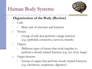
Human Body Systems. Justin Grosdidier. Table of Contents. Digestive System- Slides 3-13 Circulatory System- Slides 14-24 Respiratory System- Slides 25-34 Immune System- Slides 35-44 Excretory System- Slides 45-52. The Digestive System. Function.

E N D
Human Body Systems Justin Grosdidier
Table of Contents • Digestive System- Slides 3-13 • Circulatory System- Slides 14-24 • Respiratory System- Slides 25-34 • Immune System- Slides 35-44 • Excretory System- Slides 45-52
Function • Made up of the alimentary canal and several accessory organs that break down and absorb food • When food is taken in, it isn’t in a form the body can use. The digestive system changes this food into a form that can be used and absorbed by the body. • Involves mixing food with digestive juices that break down large molecules into smaller ones
Alimentary Organs • Mouth- The teeth grind food while saliva and the enzyme salivary amylase begin breaking down carbs • Esophagus- Muscular tube the takes food from the mouth to the stomach • Stomach- Muscular pouch that receives food, mixes it with different enzymes, digests proteins, and sends chyme to the small intestine. • Small intestine- Long tube that is the main site of digestion and absorption. It is divided into 3 parts: Duodenum, Jejunum, and Ileum • Large Intestine- Completes any absorption and processes waste into feces.
Accessory Organs • Pancreas- Secretes pancreatic juice, a liquid with enzymes and sodium bicarbonate that is able to stop the digestive process of pepsin • Liver- Secretes bile; regulates blood sugar levels; metabolizes proteins, carbs, and fats; stores glycogen; recycles used up red blood cells • Gallbladder- Stores bile produced by the liver
Digestion of Large Molecules • Digestion is a system of continuously breaking down larger molecules into smaller ones • Very large food is broken down by teeth and saliva in the mouth • Enzymes in the stomach and intestines then break down food molecules into forms that can be absorbed by the body
Enzymes • Enzymes are used in digestion by breaking down macromolecules into smaller, easier absorbed molecules • Different enzymes are used to digest different types of molecules • Salivary amylase is found in saliva and digests carbs • Pepsin is found in the stomach and breaks down proteins • Pancreatic juices break down DNA, RNA, Polypeptides, and fat molecules • Bile also breaks down fats
Physical vs. Chemical Digestion • Physical or mechanical digestion is done mainly in the mouth. The teeth cut and grind food to make it easier to swallow and increase its surface area. Salivary glands begin to secrete saliva into the mouth • Chemical digestion is the breaking down of macromolecules of food into smaller molecules that are ready for absorption. This is done in the stomach and intestines by enzymes and other digestive juices
Carb and Protein Digestion • Carbohydrates are broken down by saliva, pancreatic juices, and in the lining of the small intestine. • Starch is converted first to maltose by saliva and pancreatic juice then to glucose in the lining of the SI • Proteins are mainly digested in the stomach by Pepsin. It is then completed in the SI. • This is very complicated because proteins are very large molecules that must be broken down into small amino acids which can be absorbed into the blood
System Disorders • Acid Reflux- condition in which the stomach contents leak back into the esophagus. This can cause heartburn and many other uncomfortable symptoms • Appendicitis- Inflammation and infection of the appendix and if it is left untreated the appendix can burst causing infection and even death
Sources • http://digestive.niddk.nih.gov/ddiseases/pubs/yrdd/ • AP Book • http://hepatitis.about.com/od/overview/ig/Organs-of-Digestive-System/Digestive-System-Organs.htm • http://www.nutristrategy.com/digestion.htm • http://www.rush.edu/rumc/page-1098987321204.html
Function • The circulatory system is the major means of transportation throughout the body • It transports Oxygen from the lungs to the cells of the body and CO2 from the cells to the lungs. • Moves other substances throughout the body such as nutrients absorbed by digestion • Moves hormones throughout the body • Contains cells to fight disease • Stabilizes pH and ionic concentrations of the blood • Helps regulate body temperature
Blood Vessels • Blood vessels contain a central lumen lined with an endothelium with a smooth surface to reduce resistance to blood flow • The tissue surrounding differs between the different vessels depending on the function • Capillaries are very tiny with thin walls. This organization facilitates gas exchange. • Arteries and veins have a more complex structure. They both have an outer layer of connective elastic fibers and a middle layer of smooth muscle. Arteries have a much thicker wall than that of veins. • Arteries are used to transport blood from the heart to the different body parts and veins take the blood back to the heart. • Capillaries located between the arteries and veins. They are where gas exchange takes place.
Route of the blood • Contraction of right ventricle pumps blood to the lungs via the pulmonary arteries • It then receives oxygen from the lungs and returns via the pulmonary veins to the left atrium • It then flows into the left ventricle where the oxygen-rich blood is pumped out through the aorta to the body tissue • Blood the moves from the aorta to the branches of arteries which branch into arterioles and then to the capillaries where gas exchange takes place • The capillaries rejoin forming venules which take blood to the veins. • Oxygen-poor blood enters the heart via the vena cava veins which take blood to the right atrium and then to the right ventricle where it is pumped back to the lungs
Blood • Blood is a liquid connective tissue • The cells of the blood are suspended in a watery matrix called plasma which makes up over half the blood’s volume • There are several components of blood including erythrocytes, leukocytes, and platelets • Erythrocytes- Red blood cells are the most numerous of the blood cells. Their main function is to transport oxygen and carbon dioxide • Leukocytes- White blood cells are present to fight infection and other invaders of the body • Platelets- these are fragments of bone marrow cells that are responsible for blood clotting
Erythrocytes • Red blood cells have the responsibility of transporting oxygen to the different parts of the body • They are very numerous in the blood • Everything about their structure is to increase the efficiency of gas transport • They are small disks that are biconcave to increase surface area and increase the rate of diffusion • They do not have nuclei to leave more space for hemoglobin • Each erythrocyte contains about 250 million molecules of hemoglobin so they can transport about 1 billion O2 molecules
Open vs. Closed Systems • These are the two main types of circulation of animals with many cell layers • In open circulation, circulatory fluid, hemolymph, bathes the organs directly • Contraction of the heart pumps hemolymph through vessels into connected sinuses where exchange occurs. Relaxation of the heart then draws hemolymph • In a closed system, blood is confined to vessels and is separated from the interstitial fluid. The heart pumps blood into large vessels that branch out into smaller ones that move through the organs. • Open systems are used by arthropods and many mollusks while closed systems are found in annelids, cephalopods, and all vertebrates
Circulation in Different Animals • Fish- use single circulation where the heart has an atrium and a ventricle and the blood passes through the heart once in each circuit • Amphibians- three-chambered heart: two atria and one ventricle. A ridge in the ventricle diverts most of the oxygen-poor blood into the pulmocutaneous circuit and most of the oxygen-rich blood into the systemic circuit. This changes when underwater. • Reptiles- three-chambered heart with septum partially dividing the ventricle. In crocodilians it is complete but the circuits are connected where the arteries exit the heart. • Mammals- four-chambered heart in which the left side receives and pumps oxygen-rich blood and the right receives and pumps oxygen-poor blood
Disorders • Arrhythmia- This is an abnormal beating of the heart. Usually this means the heart beats abnormally faster or slower. This is usually congenital and caused by a heart defect. Medication, surgery, and pacemakers are the normal treatment for arrhythmias • Hypertension- This is also known as high blood pressure. Hypertension is very common. It increases the risk of heart attack and other heart diseases.
Sources • AP Book • http://faculty.clintoncc.suny.edu/faculty/michael.gregory/files/bio%20102/bio%20102%20lectures/circulatory%20system/circulat.htm • http://users.rcn.com/jkimball.ma.ultranet/BiologyPages/B/Blood.html
Function • The Respiratory system is a group of organs and tissues that allows us to breathe • This is the system that is responsible for bringing oxygen into the body and removing waste carbon dioxide • The main parts are the airways, lungs, and connected blood vessels
Alveoli • The alveoli are small air sacs that are clustered at the tips of the smallest bronchioles within the lungs. • This is the portion of the lung where the gas exchange occurs • The human lung contains millions of alveoli, which together have a surface area of about 100 m2, fifty times the area of the skin • Oxygen diffuses across the epithelium and into a web of capillaries while carbon dioxide diffuses from the capillaries into the air space
CO2 and O2 Transport • Oxygen is taken in via the air we breathe • It travels in through the trachea, to the bronchi, bronchioles, and finally to the alveoli • It is then diffused into the blood through capillaries which go back to the heart • Erythrocytes in the blood attach to and carry up to a billion oxygen molecules per cell • From the heart, oxygen-rich blood is pumped to the various parts of the body • When the blood reaches it’s destination, it swaps oxygen with carbon dioxide which is taken back to the heart • From the heart this oxygen-poor blood is pumped to the lungs where the CO2 diffuses into the alveoli and leaves the body when we exhale
Inhalation vs. Exhalation • Mammals use what is called Negative Pressure Breathing which pulls instead of pushes air into the lungs • This is possible because of the diaphragm • When the diaphragm contracts and moves down it creates a negative pressure area and pulls air into the lungs (Inhalation) • Then as the diaphragm relaxes, the pressure becomes greater inside the body so the air exits the lungs and out the airway (Exhalation)
Disorders • Asthma- This is a serious disorder involving the narrowing of the bronchial airways and restricts airflow into and out of the lungs. This causes coughing, wheezing, and difficulty in breathing • Pneumonia- This is inflammation of the lungs usually caused by bacteria. The alveoli will become filled with fluid and inflammatory cells making the lung solid and restricting space to take in air.
Sources • AP Book • http://www.nhlbi.nih.gov/health/dci/Diseases/hlw/hlw_respsys.html • http://www.ivy-rose.co.uk/HumanBody/Respiratory/Respiratory_Conditions.php
Function • The immune system is the defense mechanism of the body. • It enables an animal to avoid or limit harmful infections by either preventing or removing harmful invaders from the body • It works to recognize and identify pathogens that may be invading the body
Major Organs • Skin-outer protection • Bone marrow- produces blood cells • Thymus- produces mature T cells • Spleen- filters the blood • Lymph nodes- filter the fluid, lymph
Antigens and Antibodies • Any foreign substance that is recognized by lymphocytes and triggers a response is called an antigen • Usually polysaccharides or proteins • Are either secreted into the extracellular fluid or protrude from the surface of pathogens or other foreign cells • These are what the white-blood cells recognize and what triggers the immune response. • When the body fights of a certain pathogen the B cells will sometimes produce plasma cells that secrete a soluble form of the antigen receptor • These are called antibodies and the help the body to be more efficient in recognizing and reacting to familiar pathogens
Innate vs. Acquired Immunity • The innate immune response is the first line of defense • It consists of the exterior barriers (skin), TLR, Antimicrobial peptides and proteins, inflammatory responses, and natural killer cells • Acquired immunity is slower to develop • This is the recognition of familiar pathogens by the B and T cells • They recognize antigens produced by pathogens • They also produce antibodies which help the recognition process
Humoral vs. Cell-Mediated • Humoral immunity is an immune response as a result of antibodies secreted by B Lymphocytes • Antibodies bond to intruding antigens and mark them for destruction and removal • Cell-Mediated immunity is the immune response that involves macrophages, natural-killers, T Lymphocytes, and several cytokines • This is the opposite of humoral immunity as it doesn’t involve any use of antibodies
B and T Lymphocytes • Both B and T cells start out in the bone marrow but T cells then migrate to the thymus where they mature while B cells mature in the bone marrow • “T” for Thymus/ “B” for Bone Marrow • These are the cells required for acquired immunity as they recognize and inactivate foreign cells and molecules • These cells have antigen receptors that they use to recognize antigens given off by pathogens invading the body • B cells can produce antibodies that help recognize antigens
Antibiotics and Bacteria • Antibiotic- “Against living things” • Sometimes our immune system needs a little help in the battle against bacteria • If you are diagnosed with a bacterial infection you will usually be given some antibiotics • Antibiotics are mainly used for killing or preventing the growth of bacteria • They are very useful but are ineffective against viral or fungal infections, however
Disorders • Sometimes, the immune system will turn against particular molecules of the body itself causing an autoimmune disease. • There are many forms of autoimmune diseases. One disease, Lupus, causes the body to create antibodies against histones and DNA released by breakdown of body cells. • This causes skin rashes, fever, arthritis, and kidney dysfunction. • HIV, the pathogen that causes AIDS, both escapes and attacks the acquired immune response. • HIV infects helper T cells and mutates at a high rate making it impossible to get rid of • Over time, HIV infection destroys the immune system by causing the loss of T cells • HIV is incurable, but some treatments can work to slow HIV reproduction and progression to AIDS
Sources • AP Textbook • http://www.thebody.com/content/art1788.html • http://en.wikipedia.org/wiki/Cell-mediated_immunity • http://www.bacteriamuseum.org/cms/How-We-Fight-Bacteria/antibiotics.html
Function • The excretory system has the duty of emptying the body of nitrogenous and other wastes • It also regulates water and ion levels in body fluids
Nitrogenous wastes • Ammonia can only be tolerated at low concentrations so animals need access to lots of water • Most common in aquatic species • Mammals, most amphibians, sharks, and some bony fishes and turtles excrete urea • This is the product produced in the liver that combines ammonia with carbon dioxide • Very low toxicity but has a high energy cost • Insects, land snails, many reptiles, and birds excrete Uric Acid • This is relatively nontoxic and doesn’t readily dissolve in water • Can be secreted as a semisolid paste with little water loss
Excretory Processes • Filtration: Excretory tubule collects a filtrate from the blood. Water and solutes are forced by blood pressure across the membranes of a cluster of capillaries • Reabsorption: The transport epithelium reclaims valuable substances from the filtrate and returns them to the body fluids. • Secretion: Other substances, such as toxins and excess ions, are extracted from body fluids and added to the contents of the excretory tubule. • Excretion: The altered filtrate (urine) leaves the system and the body.
Disorders • Gout is a disorder where a human accumulates more than the usual amount of uric acid • Since it’s not water-soluble the acid is stored in the body, usually in the joints, causing pain and deformation of the joints • Kidney stones affect millions of people around the world • They can be a variety of sizes from microscopic to filling the entire renal pelvis • As the stone passes down the ureter the person experiences much pain and the kidney may be ineffective for a period of time