Download
1 / 7
70 likes | 270 Views
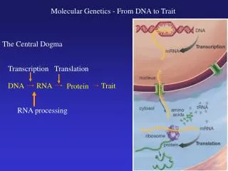
DNA. Timeline to the discovery of DNA: 1928 – Fredrick Griffith discovers non-virulent bacteria ( Streptococcus pneumoniae ) become virulent when in contact with dead pathogenic bacteria calls the process transformation

E N D
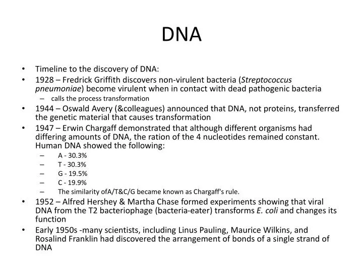
DNA • Timeline to the discovery of DNA: • 1928 – Fredrick Griffith discovers non-virulent bacteria (Streptococcus pneumoniae) become virulent when in contact with dead pathogenic bacteria • calls the process transformation • 1944 – Oswald Avery (&colleagues) announced that DNA, not proteins, transferred the genetic material that causes transformation • 1947 – Erwin Chargaff demonstrated that although different organisms had differing amounts of DNA, the ration of the 4 nucleotides remained constant. Human DNA showed the following: • A - 30.3% • T - 30.3% • G - 19.5% • C - 19.9% • The similarity ofA/T&C/G became known as Chargaff's rule. • 1952 – Alfred Hershey & Martha Chase formed experiments showing that viral DNA from the T2 bacteriophage (bacteria-eater) transforms E. coli and changes its function • Early 1950s -many scientists, including Linus Pauling, Maurice Wilkins, and Rosalind Franklin had discovered the arrangement of bonds of a single strand of DNA
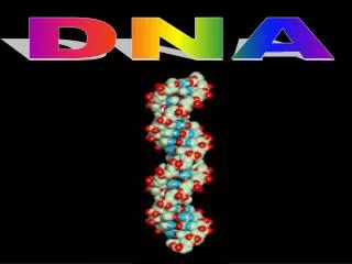
Double Helix In 1953, James Watson and Francis Crick discovered the structure of DNA .The discovery would not have been possible, though, without the work of Rosalind Franklin. Franklin's image of DNA using X-ray crystallography allowed Watson to calculate out the size and structure of the double helix of DNA. • From this picture Watson & Crick assembled their model of double helix model of DNA with the following properties: • both strands are anti-parrallel • 5' to 3' • moving in opposite directions • the sugar backbone (deoxyribose) resides on the outside • nucleotides are on the inside and paired in the following function • purines (A & G) are attached to pyrimidines (C & T) • A is attached to T with two hydrogen bonds • C is attached to G with three hydrogen bonds • explained the basis for Chargaff's rule
DNA Replication • DNA Replication is a semiconservative process where the new DNA is copied onto a parental (conserved) strand. It takes place with surprising efficiency and speed copying ~10 billion base pairs in a few hours with little or no errors. • Origin of replication: site of initiation of replication • bacteria have a single site while Eukaryotes have multiple sites • proteins recognize site and open up a replication bubble • as replication begins a replication forks form as replication proceeds in both directions • nucleoside triphosphates are added 1 at a time by DNA polymerase (~50/sec) in the 5' to 3' direction (copied 3' to 5') • energy powering exergonic process comes from cleaving two of the 3 Pi from the molecule • replication forks eventually fuse completing the newly formed strands
Antiparallel elongation • Antiparallel elongation • since nucleotides can only be added to the 3' end of the newly forming strand, different mechanisms must be in place for the antiparallel strand • leading strand - 3' to 5' • an RNA primer (5-10 nucleoside long fragment) is needed for attachment of DNA pol III • RNA attached with the enzyme primase • DNA polymerase III attaches to the primer and adds nucleosides one at a time in the 5' to 3' direction • replication continues until completion or meeting another replication fork • lagging strand - 5' to 3' • DNA pol III attaches at the replication fork and copies back to the growing strand in the 5' to 3' direction is small 100 to 200 nucleotide segments called Okazaki fragments • replication continues until DNA pol III reaches a primer then falls off • DNA pol I replaces the RNA primer with DNA • Okazaki fragments are joined (ligated) by DNA ligase as DNA pol I detaches
Other proteins involved • helicase- unwinds the double helix for replication at the replication fork • topoisomerase - relieves supercoiling caused by helicase • single-strand binding protein - stabilizes the DNA strand that has been unwound until it is replicated
Telomeres • Small sections of DNA at the 3' end of the DNA cannot be replicated as the RNA primer occupies the space. As a result daughter chromosomes are shorter that the parent chromosomes. • telomeres are regions of DNA located at the ends of chromosomes • contain 100 - 1000 repeating units (TTAGGG) • protect internal gene sequences from erosion • get shorter with each replication • associated with the aging process • telomerase is an enzyme active in germ cells and restores the length to the chromosomes • is inactive in somatic cells • may protect somatic cells from cancer
DNA Repair mechanisms • Although the polymerases check the nucleotides for accuracy as they are added, 1 in 100,000 base pairs ends in error. • Mismatch repair - nucleotide excision repair • incorrect nucleotide is excised with nuclease (protein that cuts DNA) • DNA polymerase fills in the sequence • ligase connects the strands • xerodermapigmentosum is a disease caused by a mutation in the excision repair mechanism which causes the person to be susceptible to skin damage by the sun