Download
1 / 58
580 likes | 688 Views
Case Studies. St. Jude Medical. Single Chamber ECG Analysis. ECG #1. Programmed Parameters Mode………………………………………….. VVI Base Rate……………………………………….. 70 ppm Magnet Response…………………….. Battery Test Hysteresis Rate………………………………… Off ppm. T Temporary programmed value. 1.0 Second.

E N D
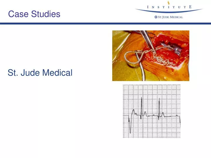
Case Studies St. Jude Medical
Single Chamber ECG Analysis ECG #1 Programmed Parameters Mode………………………………………….. VVI Base Rate……………………………………….. 70 ppm Magnet Response…………………….. Battery Test Hysteresis Rate………………………………… Off ppm T Temporary programmed value 1.0 Second 7 Mar 2000 23:20
VVI Normal Capture and Sensing Single Chamber ECG Analysis Answer ECG #1
Single Chamber ECG Analysis ECG #2
VVI Normal Capture and Sensing with initiation of Hysteresis Single Chamber ECG Analysis Answer ECG #2
Single Chamber ECG Analysis ECG #3
VVI Loss of Ventricular Sensing Single Chamber ECG Analysis Answer ECG #3
Single Chamber ECG Analysis ECG #4 1.0 Second
Single Chamber ECG Analysis ECG #4
VVI Normal Capture and Sensing Single Chamber ECG Analysis Answer ECG #4
Single Chamber ECG Analysis ECG #5
Single Chamber ECG Analysis ECG #5
VVI Normal Capture Ventricular Undersensing Single Chamber ECG Analysis Answer ECG #5
Single Chamber ECG Analysis ECG #6
Single Chamber ECG Analysis ECG #6
VVI Loss of Ventricular Capture Normal Sensing Single Chamber ECG Analysis Answer ECG #6
Dual Chamber ECG Analysis ECG #1 Base Rate 60 ppm MTR 120 ppm AVD 200 ms PVARP 250 ms
Loss of Atrial Capture Normal Atrial Sensing Normal Ventricular Capture Ventricular Sensing Unknown Dual Chamber ECG Analysis Answer ECG #1
Dual Chamber ECG Analysis ECG #2 Base Rate 60 ppm MTR 120 ppm AV 200 ms PV 150 ms Min. PV 75 ms PVARP 250 ms
Normal Atrial Capture Normal Atrial Sensing Normal Ventricular Capture Ventricular Sensing Unknown Dual Chamber ECG Analysis Answer ECG #2
Dual Chamber ECG Analysis ECG #3 Base Rate 60 ppm MTR 120 ppm AV 200 ms PV 150 ms PVARP 250 ms
Normal Atrial Capture Possible Psuedofusion on 4th atrial output Atrial Sensing Unknown Loss of Ventricular Capture Normal Ventricular Sensing Functional Loss of Ventricular Sensing Dual Chamber ECG Analysis Answer ECG #3
Dual Chamber ECG Analysis ECG #4 Base Rate 60 ppm MTR 120 ppm AV 200 ms PV 200 ms PVARP 250 ms
Normal Atrial Capture Atrial fusion on 3rd atrial output Normal Atrial Sensing Normal Ventricular Capture Normal Ventricular Sensing Dual Chamber ECG Analysis Answer ECG #4
Dual Chamber ECG Analysis ECG #5 Base Rate 60 ppm MTR 120 ppm AV 200 ms PV 200 ms PVARP 250 ms
Normal Atrial Capture Atrial Sensing Unknown Normal Ventricular Capture Fusion on 2nd ventricular output Normal Ventricular Sensing Dual Chamber ECG Analysis Answer ECG #5
Dual Chamber ECG Analysis ECG #6 Base Rate 60 ppm MTR 120 ppm AV 200 ms PV 200 ms PVARP 250 ms
Normal Atrial Capture Atrial Sensing Unknown Normal Ventricular Capture Fusion on 2nd ventricular output Ventricular Sensing Unknown Dual Chamber ECG Analysis Answer ECG #6
Dual Chamber ECG Analysis ECG #7 Base Rate 60 ppm MTR 120 ppm AV 200 ms PV 200 ms PVARP 250 ms
Normal Atrial Capture Normal Atrial Sensing Normal Ventricular Capture Normal Ventricular Sensing Dual Chamber ECG Analysis Answer ECG #7
Dual Chamber ECG Analysis ECG #8 Base Rate 60 ppm MTR 120 ppm AV 200 ms PV 200 ms PVARP 250 ms
Normal Atrial Capture with one beat showing functional loss of atrial capture Atrial Undersensing Normal Ventricular Capture Ventricular Sensing Unknown Dual Chamber ECG Analysis Answer ECG #8
Dual Chamber ECG Analysis ECG #9 Base Rate 60 ppm MTR 120 ppm AV 200 ms PV 200 ms PVARP 250 ms
Normal Atrial Capture Normal Atrial Sensing Normal Ventricular Capture Normal Ventricular Sensing Dual Chamber ECG Analysis Answer ECG #9
Dual Chamber ECG Analysis ECG #10 Base Rate 60 ppm MTR 120 ppm AV 200 ms PV 200 ms PVARP 250 ms
Normal Atrial Capture Normal Atrial Sensing Normal Ventricular Capture with two beats of functional loss of capture Ventricular Undersensing Dual Chamber ECG Analysis Answer ECG #10
Dual Chamber ECG Analysis ECG #11 Base Rate 60 ppm MTR 120 ppm AV 150 ms PV 150 ms PVARP 250 ms
Atrial Capture Unknown Normal Atrial Sensing Normal Ventricular Capture Ventricular Sensing Unknown Dual Chamber ECG Analysis Answer ECG #11
Dual Chamber ECG Analysis ECG #12 Base Rate 60 ppm MTR 120 ppm AV 200 ms PV 150 ms Min. PV 88 ms PVARP 250 ms
Normal Atrial Capture Normal Atrial Sensing Normal Ventricular Capture Ventricular Sensing Unknown Initiation of a Pacemaker Mediated Tachycardia (PMT) with following a PVC Dual Chamber ECG Analysis Answer ECG #12
Dual Chamber ECG Analysis ECG #13 Base Rate 60 ppm MTR 120 ppm AV 200 ms PV 150 ms Min. PV 88 ms PVARP 250 ms
Loss of Atrial Capture initiating a Pacemaker Mediated Tachycardia (PMT) Normal Atrial Sensing Normal Ventricular Capture Ventricular Sensing Unknown Dual Chamber ECG Analysis Answer ECG #13
Dual Chamber ECG Analysis ECG #14 Base Rate 60 ppm MTR 120 ppm AV 200 ms PV 150 ms Min. PV 88 ms PVARP 250 ms
Normal Atrial Capture Atrial Sensing Unknown Normal Ventricular Capture Normal Ventricular Sensing The retrograde P-wave after the PVC is not seen because it falls in PVARP just like it should Dual Chamber ECG Analysis Answer ECG #14
ICD ECG Analysis ECG #1
ICD ECG Analysis ECG #1
ICD ECG Analysis Answer ECG #1 • T-Wave sensing • longer decay delay • Threshold start higher
Twiddler‘s Syndrome Presented with left hemi-diaphragmatic stimulation from atrial lead
Twiddler‘s Syndrome Courtesy of Dr. F. Venditti, Lahey Clinic, MA`