Download
1 / 11
110 likes | 114 Views
This study investigates the effect of Poly-L-Lysine (PLL) substrates on the ex vivo expansion and erythroid differentiation of human hematopoietic stem cells (HSCs). Results show that PLL coatings promote HSC proliferation and do not alter their stemness markers. Higher PLL concentrations enhance the expansion of CD34+ cells and increase erythroid differentiation and enucleation. These findings suggest that PLL may be an important material for enhancing HSC expansion and erythroid differentiation.

E N D
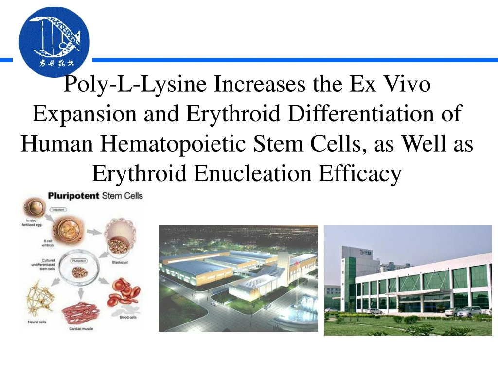
Poly-L-Lysine Increases the Ex Vivo Expansionand Erythroid Differentiation of Human HematopoieticStem Cells, as Well asErythroid Enucleation Efficacy
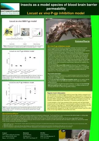
Ex vivo expansion and differentiation of HSCson PLL substrates
Tissue culture plates were coated with various concentrationsof PLL (0.001, 0.01, and 0.1 weight%), and theoxygen, nitrogen, and carbon contents on the coated surfaceswere quantified by X-rayphotoelectron spectroscopy amine groups
FIG. 1. Poly-L-lysine (PLL) substrates (0.001%, 0.01%, and 0.1%) enhance the ex vivo expansion of hematopoieticstem cells (HSCs). Ex vivo expansion was analyzed by cellcounting (A), cell division assay of CD34+ cells (B), andfluorescence-activated cell sorting analysis of CD34+ Lin -cells (C) at day 1 and 3. These results demonstrate that PLL, at acoating concentration of 0.01% or more, increased the totalcell number of CD34+ cells through an increase in celldivision, without altering the percentage of HSCs with theCD34+ Lin - surface marker expression pattern or the differentiationpotential of CD34+ cells. FIG. 2. The PLL substrate (0.01%) does not alter the multipotency of HSCs. The total colony number (A) and thepercentage of various types of colonies (B) were observed at 14 days in culture using the colony-forming assay.
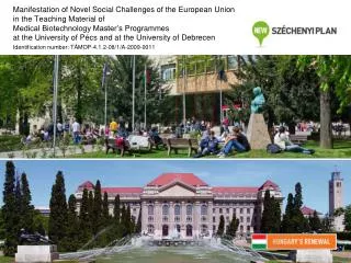
Erythroid differentiation and RBC productionon PLL substrates
These results suggest that the 0.01%PLL substrate enhanced the expansion of differentiatingHSCs at early but not late phases. 0-7 7-13 13-17 17-21 FIG. 3. PLL substrates (0.001%, 0.01%, and 0.1%) increaseerythroid differentiation of CD34+ cells. The foldincrease of erythroid cells (A), the percentage of CD71+GPA+ cells (B) and enucleated cells (C) for 21 days areshown. GPA+SYTO64- cells were regarded as enucleatedred blood cells. CD71 and Glycophorin A (GPA) are cellsurface markers of erythroid cells. These resultssuggests that the PLLsubstrates mainly induced enucleation of erythroid cells atphase III rather than at phase IV
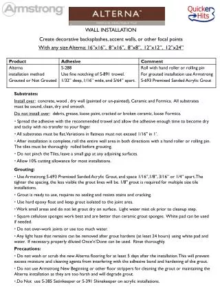
More FIG. 4. Effect of the PLL substrate(0.01%) on erythroid differentiation, asshown bybenzidine staining (A), RT-PCRwith erythropoiesis-related primers (B) andOxygen-carry abilities (C) of hemoglobin in the erythroid cells at 17 days.Black arrowheads indicate benzidine+ /hematoxylin-cells as enucleated erythrocytes.Oxygen equilibrium curves weredetermined using an automated apparatus.CB and PB indicate umbilical cord bloodand adult peripheral blood, respectively. Theseanalyses confirmed that the 0.01% PLL substrate enhancedthe enucleation of erythroid cells and, in addition, elevated theexpression of erythropoiesis-related genes during erythroid differentiation. PLL substrate is able to enhanceoxygen-carrying ability of erythroid cells.
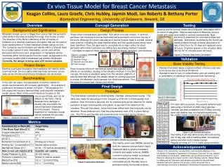
Enucleation efficacy and direction of enucleationon the 0.01% PLL substrate
The interaction between HSCs and the PLL substrate (0.01%) stimulates downward enucleation. This result strongly supports the fact that the PLLsubstrate is able to increase PI3K activity and cellular polarizationof erythroid cells followed by an increase inenucleation. phosphoinositide 3-kinase (PI3K) activity of erythroid cells was visualized by anti-PIP3 (green) and DAPI (blue) Enucleating cells were observed via confocal imaging in Z-section by using ananti-GPA antibody (green), phalloidin (red), and DAPI (blue, left panel), concluding which an enucleation mechanisticmodel is also shown (right panel)
Conclusions 1. PLL substrates increased the totalnumber of HSCs without altering their expression of surfacemarkers related to stemness. 2. Higher coatingconcentrations of PLL had a greater influence on the ex vivoexpansion of CD34+ cells, with no change in the percentageof CD34+ Lin - cells. 3. The number of cells thatunderwent erythroid differentiation followed by enucleationincreased on PLL substrate versus the control. 4. These results indicate that PLL might serve as an importantmaterial to enhance the ex vivo expansion and erythroiddifferentiation of HSCs, and especially theenucleation of erythroid cells to form RBCs.