Download
1 / 48
480 likes | 656 Views
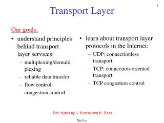
Membrane Transport “Pores, Porters and Pumps”. CH353 February 26-28, 2008. Summary. Thermodynamics and Kinetics of Membrane Transport Classification of Membrane Transport Proteins Channels, Porters, Primary Active Transporters Primary Active Transporters

E N D
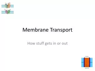
Membrane Transport“Pores, Porters and Pumps” CH353 February 26-28, 2008
Summary • Thermodynamics and Kinetics of Membrane Transport • Classification of Membrane Transport Proteins • Channels, Porters, Primary Active Transporters • Primary Active Transporters • driven by hydrolysis of phosphoanhydride bonds • Porters (secondary active transport & facilitated diffusion) • driven by electrochemical potential • Systems combining active transporters and porters • Channels (for water and ions) • Regulation of ion channels • voltage and ligand gating • action potential and synaptic function
[Solute] membrane [Solute] aqueous KD x P = K = Diffusion Across Membranes Diffusion rate is proportional to permeability of solute Permeability constant (P) depends on: • Partition constant (K) of solute Urea, K = 0.0002; Diethylurea, K = 0.01 • Diffusion constant (D) of membrane • is proportional to viscosity • viscosity of membrane ~100-1000x greater than that of water • Thickness of membrane (x) 3–5 nm • K and D vary with lipid composition and position dx within membrane
dn dt dn dt KD x = A (C1aq – C2aq) Diffusion rate ( ) Diffusion Across Membranes • Thickness (x) and diffusion constant (D) are similar for most membranes • Thus diffusion across a membrane is proportional to the partition constant of the solute (K) and the difference in concentration (chemical gradient) or electrical gradient across a membrane (membrane potential, Vm) • Electrochemical gradient / potential: combination of electrical and chemical differences across a cell membrane For membrane transport: • partition constant of solute is irrelevant • depends on electrochemical gradient = AP(C1aq – C2aq)
Diffusion Accelerated by Transporters • Diffusion rate is accelerated by lowering its activation energy, ∆G‡ • Transporters lower ∆G‡, providing another path through a membrane • Facilitated Diffusion: transport down an electrochemical gradient • Transporters are like Enzymes: • Lowers ∆G‡ (faster rate) • Substrate specificity • Saturation kinetics • No effect on ∆G of process
Kinetics of Transporters Vmax α-D-glucose α-D-mannose Initial Rate of Monosaccharide Transport, V0 (mmol/min) α-D-galactose Passive Diffusion (no GLUT1) K0.5
k1 k2 Sout + T T•S complex T + Sin k-1 k-2 Vmax[S]out Kt + [S]out k2[Tt][S]out Kt + [S]out Vmax 1 + Kt /[S]out V0 = k2[T•S] = = = k2 + k-1 k1 Ktransport (Kt) = Kt is [S] at ½Vmax Kinetics of Transporters for initial reaction conditions (Sout >> Sin): assume k-2 = 0 and [T•S] is constant Kt is similar to Km; the terms K½ or K0.5 are more commonly used
∆G = ∆G′º + RT ln () + RT ln () + ZF∆y P S C2 C1 ∆G of chemical reactions S → P ∆G of concentration gradient C1→ C2 ∆G of membrane potential y1→ y2 Thermodynamics of Transport R = gas constant = 8.315 J / mol • K (1.987 cal / mol • K) T = absolute temperature (K) Z = charge of solute • number of moles (mol) [electrogenic transport] F = Faraday constant = 96,480 J / V • mol (23,060 cal / mol • K) ∆y = y 2 - y 1 = membrane potential ∆G′º + RT ln (P/S) = 0, except for Primary Active Transport ∆G = 0 at equilibrium [resting potential]
[K+]out [K+]in [K+]out [K+]in RT ln = ZF∆y RT ZF (8.315 J/mol•ºK)(310 ºK) (+1)(98,060 J/mol•V) 4 mM 140 mM ∆y = ln = -93.5 mV Resting or Equilibrium Potential Problem: • The plasma membrane of a neuron is selectively permeable to K+. • If [K+]in = 140 mM and [K+]out = 4 mM, what membrane potential is needed to balance the transport of K+ out of the cell? Solution: At equilibrium: ∆G concentration gradient = ∆G membrane potential ∆y = ln (Nernst Equation)
P S C2 C1 ∆G = ∆G′º + RT ln () + RT ln () + ZF∆y Thermodynamics of K+ Transport Group Problem • The resting potential of a neuron is actually -70 mV on the inside • What is the ∆G for transport of K+? • Which direction is K+ spontaneously transported? Assume: [K+]in = 140 mM; [K+]out = 4 mM; T = 37ºC R = 8.315 J / mol • K; F = 96,480 J / V • mol
Transporter Classification System (http://www.tcdb.org/) Classes • Channels/Pores • Electrochemical Potential-Driven Transporters • Primary Active Transporters • Group Translocators • Transport Electron Carriers • Accessory Factors involved in Transport • Incompletely Characterized Transport Systems
Transporter Classification System (http://www.tcdb.org/) • Channels/Pores 1.A.α-Type channels 1.B. β-Barrel porins 1.C. Pore-forming toxins (proteins and peptides) 1.D. Non-ribosomally synthesized channels 1.E. Hollins 1.F. Vesicle fusion pores 1.G. Paracellular channels
Transporter Classification System (http://www.tcdb.org/) • Electrochemical Potential-Driven Transporters 2.A. Porters (uniporters, symporters, antiporters) 2.B. Non-ribosomally synthesized porters 2.C. Ion gradient-driven energizers • Primary Active Transporters 3.A. P-P-bond-hydrolysis-driven transporters 3.B. Decarboxylase-driven transporters 3.C. Methyltransfer-driven transporters 3.D. Oxidoreduction-driven transporters 3.E. Light-driven transporters
Membrane Transport Systems • Channels/Pores (α-Type)Facilitated Diffusion • Non-gated • Gated (voltage, ligand, signal) • Electrochemical Potential-driven Transporters (Porters) • Uniporter Facilitated Diffusion • Antiporter • Symporter • Primary Active Transporters (P-P bond hydrolysis driven) • ABC transporter • P-ATPase • F-ATPase Co-transport against concentration gradient (Secondary Active Transport) Transport against concentration gradient
Energy from ATP hydrolysis drives transport against electrochemical gradient Transport of a solute against its gradient is powered by transport of another down its gradient Types of Transport Typically X = Na+ or H+ electrogenic transport has a net flow of charge contributing to the membrane potential; electroneutral transport does not
Primary Active Transporters (Pumps) • A-type, F-type and V-type ATPases • transport uses a rotary mechanism (multi-subunit complexes) • 3 ATPs hydrolyzed (or synthesized) per rotation • 2 to 4 H+ (or Na+) transported per ATP • P-type ATPases • transport involves phosphorylated Asp and conformation shifts • multi-domain protein has all transporter activities • 1 ATP hydrolyzed; multiple cations (co)transported per cycle • ATP-binding cassette (ABC) Transporters • each has 2 ABC and 2 transmembrane domains/subunits • transport by dimerization of ABCs and shifting of TMDs • 1-2 ATP hydrolyzed per molecule transported
F-Type and V-Type ATPases • integral (F0, V0) and peripheral (F1, V1) multi-subunit complexes • homologous hexameric ATPase complexes (α3β3 and A3B3) • homologous rotor complexes (dec12 and DFdc6) • 1 H+ carrier (Glu) per subunit; F-type transports ~2x more H+ per ATP • non-homologous a subunits but conserved mechanism • Reversible in vitro but opposite roles in vivo (opposite rotations) • F-type is ATP synthase using [H+]; V-type is H+ pump using ATP Nishi & Forgac 2002, Nat. Rev. Mol. Biol. 3:94
Vacuolar (V-type) ATPases • Structure and Activity: • ATP-hydrolyzing peripheral complex (V1) (640 kDa) • H+-translocating integral assembly (V0) (260 kDa) • 6 c subunits in rotor: maximum 2 H+ transported per ATP (higher pH gradients than for F-type ATPases) • Electrogenic transport: requires transport of anion (e.g. Cl-) • Functions: • pH regulation in organelles (lysosomes, endosomes, vacuoles) • In specialized cells (on plasma membrane) : renal acidification, bone resorption, sperm maturation, cytoplasmic pH regulation • Multiple isoforms for specialized functions
ATP ADP + Pi V-type ATPase H+ Transport Mechanism • Subunit A hydrolyzes ATP changing its conformation • This causes 120º rotation of rotor (subunits DFdc6) • Proteolipid ring of c subunits moves past subunit a, having an essential Arg (R735), and 2 hemichannels open to either cytoplasm or lumen • The Arg removes H+ from a Glu (E) on each c subunit; H+ exits to lumen • H+ from cytoplasm neutralizes the charged Glu on c subunit, allowing it to rotate into the lipid bilayer H+ E E H+ Adapted from Forgac 2007, Nat. Rev. Mol. Biol. 8:917
V-type ATPase H+ Transport Mechanism Cycle for 60º Rotation H+ from cytoplasm enters hemichannel in subunit a H+ neutralizes charge on Glu of subunit c in the proteolipid ring H+ dissociates from Arg and exits through hemichannel to lumen Essential Arg of subunit a removes H+ from Glu on subunit c Forgac 2007, Nat. Rev. Mol. Biol. 8:917
H+in EH E– EH R RH+ H+out R V-ATPase H+ Transport Reaction H+in↔ H+out (~360º cycle) H+in EH RH+ H+out E- R E- H+ exchange on c subunit (~60º cycle)
Regulation of V-type ATPases Reversible Dissociation • V1 and V0 dissociate under low glucose conditions (yeast, insects) • Aldolase may be glucose sensor • RAVE complex required for reassembly of V-ATPase • PI3K dependence in kidney cells Plasma Membrane Localization • transport of HCO3- to cytoplasm • adenylate kinase activation • cAMP synthesis • endocytosis of V-ATPase Forgac 2007, Nat. Rev. Mol. Biol. 8:917
P-type ATPases Superfamily of active transporters (ATPases) including: • Na+K+ ATPase: maintains intracellular high [K+] and low [Na+] • Ca2+ ATPase (plasma membrane): Ca2+ homeostasis (< 0.2 μM) • Ca2+ ATPase (SERCA): concentrates [Ca2+] in SR (~10 mM) • H+K+ ATPase: gastric acidification (pH ~1) • H+ ATPase: maintains membrane potential in plants and fungi Characterized by: • Reversible phosphorylation of ATPase during transport cycle • Sensitivity to phosphate analogs, e.g. vanadate • Structural homologies (sequence and 3D structure) SERCA is prototype for structure of P-type ATPases Structures for Na+K+ ATPase and H+ ATPase (Dec 2007)
P-type ATPases • 3 cytoplasmic domains: N – Nucleotide (ATP) binding P – Phosphorylation (Asp) A – Actuator (TGES motif) • multiple transmembrane helices (10) having ion binding sites and transient channels to cytoplasm and to outside of cell (or lumen) • phosphorylation and binding of nucleotides and ions result in conformational shifts causing: • opening/closing channels • changes to ion-binding affinity Kuhlbrandt 2004, Nat. Rev. Mol. Biol. 5:282
Ca2+ ATPases Sarco-endoplasmic reticulum Ca2+ ATPase (SERCA) • Pumps Ca2+ from cytoplasm to sarcoplasmic reticulum (SR) in skeletal muscle cells (induces relaxation) • [Ca2+] = 0.1 μM in resting cell; 1 μM in contracting cell; and 2 mM in SR • ~80% of integral protein in SR Plasma membrane Ca2+ ATPase • pumps Ca2+ from cytoplasm out of cell (ubiquitous) • allosterically activated by Ca2+-calmodulin • accelerates pump when [Ca 2+] is high
Transport Cycle for SERCA Overall Reaction: 2 Ca2+in + 2-3 H+out + ATP → 2 Na2+out + 2-3 H+in + ADP + Pi E1-ATP 2 Ca2+ E1~P 2 Ca2+ E2-P 2 Ca2+ ADP 2-3 H+ 2-3 H+ outside inside Pi ATP 3 Ca2+ 2 Ca2+ E1-ATP 2-3 H+ E2 2-3 H+ E2-P 2-3 H+ [Inside] K½ K½ [Outside] Ca+: 1 μM 0.1 μM high 2 mM E1 has high affinity for Ca2+ E2 has low affinity for Ca2+
Mechanism of SERCA • A-domain is connected to 3 transmembrane helices • ATP binding to N-domain causes it to tip toward the P-domain, displacing the A-domain • This opens a channel from the cytoplasm for Ca2+ entry • Phosphorylation of P-domain causes N-domain to move back, allowing A-domain to return • This occludes the bound Ca2+ • ADP is released allowing A-domain to turn into ADP binding site and bind to P- and N-domains • This opens the channel to the lumen for to Ca2+exit
Mechanism of P-Type ATPases Kuhlbrandt 2004, Nat. Rev. Mol. Biol. 5:282
Na+K+ ATPase • maintains [K+] and [Na+] in cell; pumps 3 Na+ out and 2 K+ in • electrogenic transport accounts for some of membrane potential • tetramer of α2β2 subunits with tissue specific subunits/isoforms • sensitive to ouabain, digoxin and palytoxin • α subunit has similar 3D structure and mechanism as SERCA • 3D structure shows that Na+ and K+ may have same binding sites (Olesen et al 2007, Nature 450:1036)
Transport Cycle for Na+K+ ATPase Overall Reaction: 3 Na+in + 2 K+out + ATP → 3 Na+out + 2 K+in + ADP + Pi E1-ATP 3 Na+ E1~P 3 Na+ E2-P 3 Na+ ADP 2 K+ 2 K+ outside inside Pi ATP 3 Na+ 3 Na+ E1-ATP 2 K+ E2 2 K+ E2-P 2 K+ [Inside] K½ K½ [Outside] Na+: 12 mM 0.6 mM high 145 mM K+: 140 mM high 0.2 mM 4 mM
ATP-Binding Cassette (ABC) Transporters • Superfamily of active transporters for both import and export of diverse molecules across membranes • ABC importers found only in bacteria; require additional binding protein • Each transporter has 2 transmembrane domains (TMDs) and 2 nucleotide-binding domains (NDBs) • The NDBs are conserved, interchangable structures the TMDs vary with the molecule transported • Dimerization of NBDs changes conformation of TMDs directing alternate access to either side of membrane
Structures of ABC Transporters • ABC importers: separate subunits for NBDs and TMDs • ABC exporters: single multidomain polypeptide Hollenstein et al. 2007, Curr. Opin. Struct. Biol. 17: 412
Structure of the B12 Transporter • ABC importer for vitamin B12 is tetramer of NDBs and TMDs (BtuC2D2) • requires periplasmic B12 binding protein (BtuF) • ABC exporters do not need a binding protein Locher 2004, Curr. Opin. Struct. Biol. 14: 426
NBDs of ABC Transporters • Cooperative binding and hydrolysis of ATP • 2 NBDs form head-to-tail dimers with 2 ATPs sandwiched between • ATP binding site (P) of one domain next to the hydrolysis site (P) of the other domain • NBDs have binding sites for conserved coupling helices from TMDs Hollenstein et al. 2007, Curr. Opin. Struct. Biol. 17: 412
ATP Induced Conformational Changes ModBC-A without ATP • Coupling helices of ABC transporters with ATP are closer than those without ATP Switch Model: • 2 conformations: open dimer (- ATP), closed dimer (+ ATP) • Binding of solute to TMDs activate NBDs • ATP binding provides power for transport (closed NBDs) • ATP hydrolysis restores transporter (open NBDs) Higgins & Linton 2004 Nat. Struct. Mol. Biol. 11: 918 Sav1866 with ATP analog Hollenstein et al. 2007, Curr. Opin. Struct. Biol. 17: 412
Human ABC Proteins 12 Sub-family A (ABC1) – lipid transport 11 Sub-family B (MDR/TAP) – multi-drug resistance / T-cell antigen processing 13 Sub-family C (CFTR/MRP) – cystic fibrosis transmembrane conductance regulator / multiple resistance pump 4 Sub-family D (ALD) – peroxisomal fatty acyl-CoA 1 Sub-family E (OABP) 3 Sub-family F (GCN20) 8 Sub-family G (WHITE) – eye pigment, cholesterol
Major Facilitator Superfamily • Largest group of porters (>5000 in all kingdoms, 54 in human) • Diverse in function (uniporters, symporters, antiporters) • Most have 12 transmembrane helices (some with 14 and 24) • Low sequence homology but similar predicted topology Examples • Sugar Porter Family (2.A.1.1) • Glucose transporters (human) GLUT1 – GLUT12 [Uniporters] • Organophosphate:Pi Antiporter Family (2.A.1.4) • Glycerol- Phosphate transporter (E. coli) GlyT [Antiporter] • Oligosaccharide:H+ Symporter Family (2.A.1.5) • Lactose permease (E. coli) lacY [Symporter]
S • T1 S • T2 2 Kinetic Model Sout Sin 3 1 4 T1 T2 Model for Glucose Transport by GLUT1 • Transporter has 2 conformations • T1 facing outside; T2 facing inside • Transport of glucose proceeds by alternate access model (rocker switch) • Rate limiting step: T1↔ T2(step 4) • demonstrated using labeled glucose
Properties of Glucose Transporters GLUT1 GLUT4 GLUT2 Initial Rate / Maximum Rate, V0 / Vmax Physiological Range of Blood [Glucose]
Insulin Regulation of GLUT4-Mediated Glucose Transport in Muscle Cells • Insulin increases rate of glucose transport ~15 x
Structures of MFS Porters from E. coli Alternating Access Model – “Rocker Switch” Mechanism Locher et al. 2003, Science 301: 603
Lactose Transport in E. coli • Lactose permease lacY uses electrochemical H+ gradient for symport of lactose (secondary active transport) • H+ gradient is generated by oxidative respiration (electron transport) • Import of lactose is sensitive electron transport inhibitors
Inhibiting Secondary Active Transport of Lactose by lacY Mutants or Cyanide • Glu325 and Arg302 are both essential for coupling transport of H+ and lactose • lacY mutants are active in facilitated diffusion but not secondary active transport • Collapse of H+ electrochemical gradient produces same result • High intracellular lactose diffuses out when respiration is poisoned
Structure of Lactose Permease and Proposed Transport Mechanism • 3D structure of lactose permease with bound substrate (red) and essential Glu325 and Arg302 (green) • protonation of amino acid side chains, e.g. Glu325 and Arg302 may change ionic interactions and switch conformations; with alternate access to cytoplasm or periplasmic space
Structure of Glycerol-3-Phosphate Transporter of E. coli 3D structure of Glycerol-3-phosphate transporter with substrate binding amino acids Arg45 and Arg269 Huang et al. 2003, Science 301: 616
Glycerol-3-Phosphate : Phosphate Antiport by Rocker Switch Mechanism • Transporter alternates between conformations facing outward (Co) and inward (Ci) • Binding phosphate or glycerol-3-phosphate draws 2 arginines together facilitating the Co↔ Ci conformation switch • Conformation changes are rate limiting and temperature dependent • Binding phosphate or glycerol-3-phosphate is temperature independent Huang et al. 2003, Science 301: 616; Law et al. 2007, Biochem 46: 12190