Download
1 / 40
410 likes | 536 Views
Tissue Organisation in the Body. A&P 1 Tutor: Eleshia Howell. Chapter 3 – from p34, Ross & Wilson: Tissues are collections of cells and cell products that perform specialised, yet limited functions according to their type.

E N D
Tissue Organisation in the Body A&P 1 Tutor: Eleshia Howell Written by Eleshia Howell for use by AoCH - WEA Hunter.(c) 2012. Not to be used without the express permission of the Author.
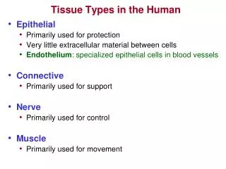
Chapter 3 – from p34, Ross & Wilson: Tissues are collections of cells and cell products that perform specialised, yet limited functions according to their type. There are four main types of tissues, each with their own sub-divisions. • Epithelial – covers the body (skin) and lines cavities, hollow organs and tubes, and is also found in Glands. • Connective – fills internal spaces, supporting other tissues; transports material; stores energy. • Muscle – specialised for contraction • Nerve – carries electrical signals from one part of the body to another. Written by Eleshia Howell for use by AoCH - WEA Hunter.(c) 2012. Not to be used without the express permission of the Author.
Epithelial Tissue • The structure of epithelial tissue is closely related to its functions, which include: • Protection of underlying structures • Secretion • Absorption • The cells are very closely packed and the intercellular substances, or matrix, is minimal. • May be simple (a single layer of cells) or stratified (several layers of cells. Written by Eleshia Howell for use by AoCH - WEA Hunter.(c) 2012. Not to be used without the express permission of the Author.
Simple Epithelium • Consists of a single layer of identical cells, sub-divided into 3 main types which are named according to their function. The more active the tissue, the taller the cells. • It is usually found on secretory surfaces. Written by Eleshia Howell for use by AoCH - WEA Hunter.(c) 2012. Not to be used without the express permission of the Author.
Squamous – composed of a single layer of flattened cells, fitting closely together like a pavement, forming a thin, smooth membrane across which diffusion occurs easily. It forms the lining of the following structures • Heart (endocardium) • Blood and lymph vessels ( endothelium) • Alveoli of the lungs • Ducts of the kidneys Cuboidal – cube shaped cells on a basement membrane, actively involved in secretion, absorption and excretion. Found in tubules of the kidneys, sweat ducts, mammary glands and some glands. Written by Eleshia Howell for use by AoCH - WEA Hunter.(c) 2012. Not to be used without the express permission of the Author.
Columnar – rectangular shaped cells, on a basement membrane, which lines many organs. Can have ancillary structures, such as microvilli and goblet cells, to enhance the function of its location. For example, columnar cells line the small intestine and have microvilli which increase the surface area of the organ, enabling an increased absorption of water and nutrients. In the trachea, the cells are ciliated (have hair like projections) which collect inhaled particles to be moved towards the throat. Stratified – consists of several layers of cells of various shapes, to help protect underlying structures from mechanical wear and tear. Sub-divided into two types: Stratified Squamous (mainly columnar, with squamous at the top where it can be shed) and Transitional (irregular, pear shaped cells allowing for stretch in the tissue). Written by Eleshia Howell for use by AoCH - WEA Hunter.(c) 2012. Not to be used without the express permission of the Author.
Connective Tissue • The most abundant tissue found in the body • Tissue cells are more widely separated and matrix is present in considerably larger amounts. • Usually fibres present in the matrix too, forming a supportive network for the cells to attach to. • Most have a good blood supply. • Four main functions: • Binding & structural support • Protection • Transport • insulation Written by Eleshia Howell for use by AoCH - WEA Hunter.(c) 2012. Not to be used without the express permission of the Author.
Connective tissue, excluding blood, is found in all organs supporting the specialised tissues. The different types of cells involved include: • Fibroblasts • Fat cells (adipocytes) • Macrophages • Leukocytes • Plasma cells • Mast cells Let’s take a look at these individually.... Written by Eleshia Howell for use by AoCH - WEA Hunter.(c) 2012. Not to be used without the express permission of the Author.
Fibroblasts Large cells with irregular processes, they produce collagen and elastic fibres. They are very active in tissue repair where they can bind together the broken surfaces of a wound, or form granulation tissue where tissue has been destroyed. Collagen fibres shrink as they grow old, resulting in adhesions. Written by Eleshia Howell for use by AoCH - WEA Hunter.(c) 2012. Not to be used without the express permission of the Author.
Fat Cells Each cell stores a single large fat droplet and can occur singly in tissue or in groups. They are especially abundant in Adipose (Fat) tissue and can vary in size / shape depending on where they are located. Written by Eleshia Howell for use by AoCH - WEA Hunter.(c) 2012. Not to be used without the express permission of the Author.
Macrophages Large irregular shaped cells that act as part of the immune system, engulfing and digesting cellular debris, foreign bodies and bacteria (phagocytosis). Some are fixed (attached) to connective tissue fibres and other roam freely. Written by Eleshia Howell for use by AoCH - WEA Hunter.(c) 2012. Not to be used without the express permission of the Author.
Leukocytes Also known as White Blood Cells (WBC’s). Normally found in small numbers in healthy connective tissue , but as they play a major role in the immune system, become more prolific during an infection. Written by Eleshia Howell for use by AoCH - WEA Hunter.(c) 2012. Not to be used without the express permission of the Author.
Mast Cells • Found in loose connective tissue, under the fibrous capsule of organs and in considerable amounts around blood vessels. • They are another aspect of the immune system response, producing tiny granules of Heparin (to reduce blood clotting) and Histamine (to reduce inflammatory reactions) to mediate the inflammation process and allergic reactions. Written by Eleshia Howell for use by AoCH - WEA Hunter.(c) 2012. Not to be used without the express permission of the Author.
Other cells found in Connective Tissue include: • Melanocytes – synthesizes and stores the brown pigment melanin. • Mesenchymal cells – stem cells that respond to injury or infection; can differentiate into various forms of cells as required, eg Fibroblasts, Macrophages etc. • Lymphocytes – specialised immune cells in Lymphoid tissue • Microphages – small phagocytic blood cells, egEosinophils, Neutrophils. Written by Eleshia Howell for use by AoCH - WEA Hunter.(c) 2012. Not to be used without the express permission of the Author.
Loose (areolar) Connective Tissue • The most generalised type of connective tissue, found almost in every part of the body, providing elasticity and tensile strength. • It connects and supports other tissues, such under the skin, between muscles, blood vessels and nerves, and the alimentary canal. • See fig 3.16 on p36 Ross & Wilson Written by Eleshia Howell for use by AoCH - WEA Hunter.(c) 2012. Not to be used without the express permission of the Author.
Adipose Tissue • Consists of fat cells in a matrix of areolar tissue. • Two types: White and Brown • White adipose tissue makes us 20-25% of body weight in a well-nourished adult. It is found supporting organs, between muscle fibres and under the skin, where it acts as insulation and an energy store. • Brown adipose tissue is present in Newborns and has a more extensive blood supply. When it metabolises, it produces less energy and more heat, helping to maintain body temperature. Written by Eleshia Howell for use by AoCH - WEA Hunter.(c) 2012. Not to be used without the express permission of the Author.
Dense Connective Tissue • Contains more fibres and fewer cells than loose connective tissue. • Two types: Fibrous and Elastic • Fibrous tissue is made up of closely packed bundles of collagen fibres with very little matrix. This kind of tissue is found in Ligaments, the Periosteum, as an outer protective layer on organs and forms muscle sheaths which extend beyond the muscle to become Tendons. • Elastic tissue is capable of considerable extension and recoil. Therefore it has fewer cells and more elastic fibres in the matrix. It is found in organs and structures where alteration of shape is required, eg: lungs, arteries, bronchi. Written by Eleshia Howell for use by AoCH - WEA Hunter.(c) 2012. Not to be used without the express permission of the Author.
Blood • Is firmer than other connective tissues. • Cells are called Chondrocytes and are less numerous, embedded in matrix reinforced by collagen and elastic fibres • There are 3 types of cartilage: • This is a fluid connective tissue and is discussed in more detail in weeks 13 & 14. Cartilage Written by Eleshia Howell for use by AoCH - WEA Hunter.(c) 2012. Not to be used without the express permission of the Author.
Hyaline Cartilage: Smooth, bluish-white tissue provides flexibility, support and smooth surfaces for bone movements. It is found – • At the end of long bones • Costal (rib) cartilage • Parts of the larynx, trachea and bronchi. Fibrocartilage: A tough, slightly flexible supportive tissue found – • Intervertebral discs • Between articulating surfaces of joints • On the rim of bony sockets, eg hip and shoulder • As Ligaments, joining bone to bone. Written by Eleshia Howell for use by AoCH - WEA Hunter.(c) 2012. Not to be used without the express permission of the Author.
Bone Elastic Fibrocartilage: Flexible tissue provides support and maintains the shape of a structure, eg ear lobe, epiglottis and internal blood vessel walls. Bone cells, called Osteocytes, are surrounded by a matrix of collagen fibres strengthened by inorganic salts, eg calcium and phosphate. This provides bone with the its strength and rigidity. Bone has considerable capacity for growth and regeneration. There are two types of Bone: Written by Eleshia Howell for use by AoCH - WEA Hunter.(c) 2012. Not to be used without the express permission of the Author.
Compact Bone – is solid, or dense in appearance. • Spongy or Cancellous Bone – has a fine, honeycomb appearance. • Bone is discussed in more detail in week 11 when we look at the Skeleton. Written by Eleshia Howell for use by AoCH - WEA Hunter.(c) 2012. Not to be used without the express permission of the Author.
Muscle tissue • Is able to contract and relax, providing movement within and of the body. • Muscle contraction requires an adequate blood supply to provide oxygen, calcium and nutrients and to remove waste products. • There are 3 types of specialised muscle cells: • Skeletal • Smooth • Cardiac Written by Eleshia Howell for use by AoCH - WEA Hunter.(c) 2012. Not to be used without the express permission of the Author.
Skeletal Muscle • Forms the muscles that move the bones of the skeleton • Are striated (striped) • Are voluntary – you can move them at will • May be consciously (e.g.writing)or sub-consciously controlled (e.g.breathing) • Fibres are cylindrical, contain several nucleii and can be up to 35cm long. • Contraction is stimulated by motor nerve impulses, originating in the brain or spinal cord, and ending at the neuromuscular junction (where nerve meets muscle tissue) Written by Eleshia Howell for use by AoCH - WEA Hunter.(c) 2012. Not to be used without the express permission of the Author.
Written by Eleshia Howell for use by AoCH - WEA Hunter.(c) 2012. Not to be used without the express permission of the Author.
Smooth Muscle • Is described as non-striated, visceral or involuntary. • Has the intrinsic ability to contract and relax, although it is usually only relaxed for very short periods of time. • Contraction is slower and more sustained. • Autonomic nerve impulses, some hormones and local metabolites stimulate contraction. • Found in the walls of hollow organs, eg: blood vessels, uterus, ducts, glands etc. • The cells are spindle shaped, with only one nucleus. • The muscle fibres are generally bundled together to form a sheet of muscle. Written by Eleshia Howell for use by AoCH - WEA Hunter.(c) 2012. Not to be used without the express permission of the Author.
Written by Eleshia Howell for use by AoCH - WEA Hunter.(c) 2012. Not to be used without the express permission of the Author.
Cardiac Muscle • Found only in the heart wall. • Involuntary • Has striations, similar to skeletal muscle • Each fibre has a nucleus and one or more branches, which is in very close contact with adjacent cells. Microscopically, these joins or intercalated discs, can be seen as lines that are thicker and darker than ordinary muscle striations. • The significance of the closely aligned structure is that it allows the wave-like contraction of a heart beat (individual cells do not need to be stimulated to contract). Written by Eleshia Howell for use by AoCH - WEA Hunter.(c) 2012. Not to be used without the express permission of the Author.
Written by Eleshia Howell for use by AoCH - WEA Hunter.(c) 2012. Not to be used without the express permission of the Author.
Nerve Tissue • Two types of tissue are found in the nervous system: • Excitable cells – called neurones, they initiate, receive, conduct and transmit information • Non-excitable cells – known as glial cells, these support the neurones. More on nerve cells will be discussed in week 4. Written by Eleshia Howell for use by AoCH - WEA Hunter.(c) 2012. Not to be used without the express permission of the Author.
Membranes • Membranes are sheets of tissue that cover or line surfaces or separate organs or parts of organs from each other. Many membranes provide specialized secretions. • The two groups of membranes we will look at are: • Epithelial Membranes – further subdivided into Mucous and Serous • Synovial Membranes – these line cavities of moveable joints and also surrounds tendons that are in close contact with bony structures. It consists of areolar connective tissue and elastic fibres. It secretes clear, sticky, oily synovial fluid which lubricates and nourishes the joints. Written by Eleshia Howell for use by AoCH - WEA Hunter.(c) 2012. Not to be used without the express permission of the Author.
Mucous Membranes – sometimes referred to as mucosa, this lines the alimentary, respiratory and genitourinary tracts. Some of the epithelial cells on the surface of a membrane are actually Goblet cells, which produce mucous – when the cell fills up with the mucous, it bursts and discharges the fluid. This is why mucous membranes appear moist and slippery; this helps to protect the membrane and in areas such as the respiratory tract, even helps to trap foreign particles from entering the lungs. Written by Eleshia Howell for use by AoCH - WEA Hunter.(c) 2012. Not to be used without the express permission of the Author.
Serous Membrane – sometimes referred to as Serosa, consist of a double layer of loose areolar connective tissue lined by simple squamous epithelium. The parietal layer lines a cavity and the visceral layer surrounds organs (viscera) within the cavity. Serous membranes secrete serous fluid, decreasing friction as the surfaces of organs glides across other tissue or organs. There are three sites where serous membranes are found: • The Pleura lining of the thoracic cavity and surrounding the lungs • The Pericardium lining the pericardial cavity and surrounding the heart • The Peritoneum lining the abdominal cavity and the surrounding the abdominal organs. Written by Eleshia Howell for use by AoCH - WEA Hunter.(c) 2012. Not to be used without the express permission of the Author.
Glands see p41 Ross & Wilsom • Groups of epithelial cells that produce specialised secretions. • Glands that secrete (directly or indirectly) through a duct are known as EXOCRINE glands • Glands that secrete into blood and lymph are called ENDOCRINE glands and the substance is called a Hormone. Written by Eleshia Howell for use by AoCH - WEA Hunter.(c) 2012. Not to be used without the express permission of the Author.
A Salivary gland – example of an exocrine gland (ducts) Written by Eleshia Howell for use by AoCH - WEA Hunter.(c) 2012. Not to be used without the express permission of the Author.
Endocrine Glands found in the Human Body (will be covered in weeks 8 and 9) Written by Eleshia Howell for use by AoCH - WEA Hunter.(c) 2012. Not to be used without the express permission of the Author.
Tissue Repair • Tissues respond to damage or injury via two processes: • Inflammation – the inflammatory response is the first response to injury. • Regeneration – begins when the initial inflammation phase is complete and any infection has been “cleaned up”. Written by Eleshia Howell for use by AoCH - WEA Hunter.(c) 2012. Not to be used without the express permission of the Author.
Inflammatory Response (p367-369 Ross & Wilson) • Can be triggered by trauma to the tissue, chemical / toxic agents or infection (invasion of pathogens). • Signs and symptoms of inflammation are: • Redness • Heat • Pain • Swelling Written by Eleshia Howell for use by AoCH - WEA Hunter.(c) 2012. Not to be used without the express permission of the Author.
Regeneration • Once phagocytosis is complete, Fibroblasts secrete collagen fibres to form scar tissue • A ‘maturation’ process begins where the collagen fibres are gradually replaced with new tissue, helped by somatic stem cells (mesenchymal cells) which produce the right type of tissue needed for the site. • Some tissue regenerates better than others – epithelia and connective tissue regenerate well, whereas cardiac and nerve tissue does not (or does so very poorly). Written by Eleshia Howell for use by AoCH - WEA Hunter.(c) 2012. Not to be used without the express permission of the Author.
Effects of Aging on Tissue Repair • The speed and efficiency of tissue repair decreases with age due to: • Slower rate of energy consumption (metabolism) • Hormones • Reduced physical activity • Altered nutritional intake • Build up of free-radical damage • The effects are: • Thinning of epithelial and connective tissues • Increased bruising and bone brittleness • Joint pain / bone fractures • Cardiovascular disease • Mental deterioration Written by Eleshia Howell for use by AoCH - WEA Hunter.(c) 2012. Not to be used without the express permission of the Author.
Summary: You should now have a general understanding of the different types of tissue present in the human body and their functions, not only on a cellular level, but for the organism as a whole. Written by Eleshia Howell for use by AoCH - WEA Hunter.(c) 2012. Not to be used without the express permission of the Author.