Download
1 / 24
240 likes | 246 Views
This lecture provides an overview of glial cells in the CNS, including their roles in myelin sheath production, blood-brain barrier maintenance, cell migration, and synaptic function. The lecture also covers the different types of glial cells and their involvement in metabolic cooperation with neurons.

E N D
M. Kálmán: The glia The lectures for EM II on Oct. 7 and 8, 2015, actualized for 2017. For the myelin sheath see materials for Semester I.
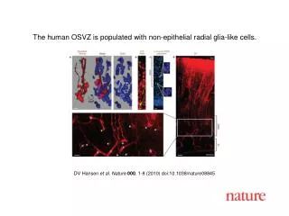
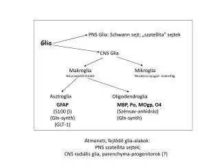
Comments to the slides • To slide 09. • Note: there are two parts of the permanent neuron production in the mature mammalian central nervous system. • In the lateral upper corner of the anterior horn of the lateral ventricle (the corner between the head of caudate nucleus and the corpus callosum) granular cells are formed in the subventricular zone for the olfactory bulb, and migrate in a glial ’channel’ along the obliterated olfactory ventricle (’rostral migratory stream’). • Granular cells are also produced in the dentate gyrus. • To Slides 10 and 11. • The components of the blood-brain barrier: • tight junctions between the endothelial cells; • extremely slow transcytosis; • P glycoprotein (pumps out lipophilic molecules); • sophisticated system of specific transports for the necessary substances int he endothelial cells and in the vascular. • Perivascular glia and pericytes maintain it. It is absent in the circumventricular organs (except for the subcommissural organ). • Other CNS barriers: • Blood-CNS fluid barrier: tight junctions between the choroidal epithelial cells. • Brain-CNS fluid barrier: tight junctions between the tanycytes of the circumventricular organs. In these two cases there are no endothelial tight junctions (see in Slide 20). • CNS fluid-extracerebral space barrier: ’neurothel’, multi-layered flat meningeal cells in the outer (subdural) part of the arachnoid. • In contrast to the former opinions, the common ependymocytes and the subpial glia limitans form no diffusion barriers. • Note: also study the slides • To Slide 12. • The axon growth and the cell migration are controlled by cell adhesion molecules, extracellular matrix molecules and humoral factors. E.g. the laminin and fibronectin promote, the tenascin and the chondroitinsulfate-containing proteoglycans inhibit the axon growth. The astrotactin supports the attachment of the migrating neurons to the radial glia, the reelin detach them. Humoral factors are e.g. the attractant netrins and the repellent semaphorins. • To Slide 14 • The term ’Tripartite’ refers to the participation of astrocytes in the synapse (’third part’ to the presynaptic and postsynaptic elements). • ’Potassium syphon’ or ’potassium drainage’ – the ’functional syncytium’ (gap junctions!) of the astrocytes distributes the potassium ions accumulated following an intense neural activity and transports into vessels the excess. • Glutamate and NH4+ uptake: The intense neuronal activity increase local glutamate concentration (transmitter release). Astrocytes inactivate glutamate forming non-transmitter glutamine with the (toxic) NH4+. • GABA – shunt: Astrocytes transform the released GABA to glutamine and return it to the GABA-ergic neurons which re-produce the GABA from it. • Glucose metabolism: Blood-borne substances are mediated by astrocytes to the neurons. Glucose can be accumulated by the astrocytes in the form of glycogen and served up to the neurons in the form of lactate. • Of course, the biochemical details are not to be memorized, leave them to Biochemistry. • To Slide 18 • At the border betwen PNS and CNS there is a an astrocyte ’mesh’. In its holed the axons pass it. At the border there is a Ranvier-node, on one side olgodendrocyte, ont he other side a Schwann-cell forms the myelin sheath. This ’histological’ border is usually not at the anatomical one. • of ‘The myelin sheath’ !