Download
1 / 5
50 likes | 127 Views
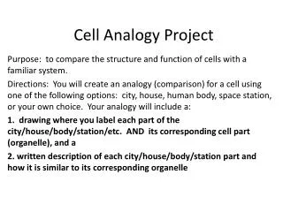
Identify the information about your cell: Location in the body, in the system (diagram needed) Function of system – in general 1. Function of cell within the system. 2. What if the cell is not functioning? D. Structure supporting function (Pictures or drawings needed)

E N D
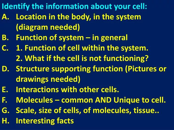
Identify the information about your cell: Location in the body, in the system (diagram needed) Function of system – in general 1. Function of cell within the system. 2. What if the cell is not functioning? D. Structure supporting function (Pictures or drawings needed) E. Interactions with other cells. F. Molecules – common AND Unique to cell. G. Scale, size of cells, of molecules, tissue.. H. Interesting facts
Suggested division of labor in team: • 1) Finds information in the texts (one or all) • 2) Slide creator: • Paraphrases into shortened texts for slides. • Identifies need for pictures and diagrams. • 3) Thinks about the flow of the show. Writes or thinks about the script.
A brush border (or striated border or brush border membrane) is the name for the microvilli-covered surface of simple cuboidal epithelium and simple columnar epithelium cells found in certain locations of the body. Microvilli are approximately 100 nanometers in diameter and their length varies from approximately 100 to 2,000 nanometers in length. Because individual microvilli are so small and are tightly packed in the brush border, individual microvilli can only be resolved using electron microscopes;[1] with a light microscope they can usually only be seen collectively as a fuzzy fringe at the surface of the epithelium. This fuzzy appearance gave rise to the term brush border, as early anatomists noted that this structure appeared very much like the bristles of a paintbrush. Brush border cells are found in two main locations: The small intestine tract: This is where absorption takes place.[2][3][4] The brush borders of the intestinal lining are the site of terminal carbohydrate digestions. The microvilli that constitute the brush border have enzymes for this final part of digestion anchored into their apical plasma membrane as integral membrane proteins. These enzymes are found near to the transporters that will then allow absorption of the digested nutrients. The kidney: Here the brush border is useful in distinguishing the proximal tubule (which possesses the brush border) from the distal tubule (which does not).[5][6] The brush border morphology increases a cell's surface area, a trait which is especially useful in absorptive cells. Cells that absorb substances need a large surface area in contact with the substance to be efficient.[7] In intestinal cells, the microvilli are referred to as striated border, while in the kidneys, microvilli are referred to as brush border.[8]
Slide Show Presentation – Criteria For 100 points: Script: accurate, flowing Ending question(s): Challenging Images – relevant, pleasing Logistics - Title slide, under 5 min. Filming - Camera is steady, looks professional, Show students’ faces, even shortly. * Creative (competition)!