Download
1 / 46
470 likes | 1.1k Views
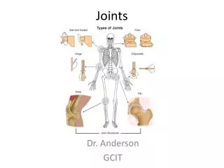
8. P A R T B. Joints. Plane Joint. Plane joints Articular surfaces are essentially flat Allow only slipping or gliding movements Only examples of nonaxial joints. Figure 8.7a. Types of Synovial Joints. Hinge joints

E N D
8 P A R T B Joints
Plane Joint • Plane joints • Articular surfaces are essentially flat • Allow only slipping or gliding movements • Only examples of nonaxial joints Figure 8.7a
Types of Synovial Joints • Hinge joints • Cylindrical projections of one bone fits into a trough-shaped surface on another • Motion is along a single plane • Uniaxial joints permit flexion and extension only • Examples: elbow and interphalangeal joints
Hinge Joints Figure 8.7b
Pivot Joints • Rounded end of one bone protrudes into a “sleeve,” or ring, composed of bone (and possibly ligaments) of another • Only uniaxial movement allowed • Examples: joint between the axis and the dens, and the proximal radioulnar joint
Pivot Joints Figure 8.7c
Condyloid or Ellipsoidal Joints • Oval articular surface of one bone fits into a complementary depression in another • Both articular surfaces are oval • Biaxial joints permit all angular motions • Examples: radiocarpal (wrist) joints, and metacarpophalangeal (knuckle) joints
Condyloid or Ellipsoidal Joints Figure 8.7d
Saddle Joints • Similar to condyloid joints but allow greater movement • Each articular surface has both a concave and a convex surface • Example: carpometacarpal joint of the thumb
Saddle Joints Figure 8.7e
Ball-and-Socket Joints • A spherical or hemispherical head of one bone articulates with a cuplike socket of another • Multiaxial joints permit the most freely moving synovial joints • Examples: shoulder and hip joints
Ball-and-Socket Joints Figure 8.7f
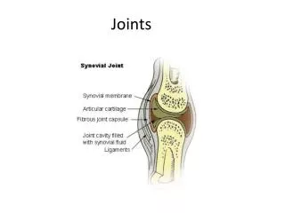
Synovial Joints: Knee • Largest and most complex joint of the body • Allows flexion, extension, and some rotation • Three joints in one surrounded by a single joint cavity • Femoropatellar joint • Lateral and medial tibiofemoral joints
Synovial Joints: Knee Ligaments and Tendons – Anterior View • Tendon of the quadriceps femoris muscle • Lateral and medial patellar retinacula • Fibular and tibial collateral ligaments • Patellar ligament Figure 8.8c
Synovial Joints: Knee – Other Supporting Structures • Anterior cruciate ligament • Posterior cruciate ligament • Medial meniscus (semilunar cartilage) • Lateral meniscus
Synovial Joints: Knee – Other Supporting Structures Figure 8.8b
Synovial Joints: Knee – Posterior Superficial View • Adductor magnus tendon • Articular capsule • Oblique popliteal ligament • Arcuate popliteal ligament • Semimembranosus tendon Figure 8.8e
Synovial Joints: Shoulder (Glenohumeral) • Ball-and-socket joint in which stability is sacrificed to obtain greater freedom of movement • Head of humerus articulates with the glenoid fossa of the scapula
Synovial Joints: Elbow • Hinge joint that allows flexion and extension only • Radius and ulna articulate with the humerus
Synovial Joints: Elbow • Annular ligament • Ulnar collateral ligament • Radial collateral ligament Figure 8.10a
Synovial Joints: Elbow Figure 8.10b
Synovial Joints: Elbow Figure 8.10d
Synovial Joints: Shoulder Stability • Weak stability is maintained by: • Thin, loose joint capsule • Four ligaments – coracohumeral, and three glenohumeral • Tendon of the long head of biceps, which travels through the intertubercular groove and secures the humerus to the glenoid cavity • Rotator cuff (four tendons) that encircles the shoulder joint and blends with the articular capsule
Synovial Joints: Shoulder Stability Figure 8.11a
Synovial Joints: Shoulder Stability Figure 8.11b
Synovial Joints: Hip (Coxal) Joint • Ball-and-socket joint • Head of the femur articulates with the acetabulum • Good range of motion, but limited by the deep socket and strong ligaments
Synovial Joints: Hip Stability • Acetabular labrum • Iliofemoral ligament • Pubofemoral ligament • Ischiofemoral ligament • Ligamentum teres Figure 8.12a
Synovial Joints: Hip Stability Figure 8.12c, d
Temporomandibular Joint (TMJ) • Mandibular condyle articulate with the temporal bone • Two types of movement • Hinge – depression and elevation of mandible • Side to side – (lateral excursion) grinding of teeth
Temporomandibular Joint Figure 8.13a, b
Sprains • The ligaments reinforcing a joint are stretched or torn • Partially torn ligaments slowly repair themselves • Completely torn ligaments require prompt surgical repair
Cartilage Injuries • The snap and pop of overstressed cartilage • Common aerobics injury • Repaired with arthroscopic surgery
Dislocations • Occur when bones are forced out of alignment • Usually accompanied by sprains, inflammation, and joint immobilization • Caused by serious falls and are common sports injuries • Subluxation – partial dislocation of a joint
Inflammatory and Degenerative Conditions • Bursitis • An inflammation of a bursa, usually caused by a blow or friction • Symptoms are pain and swelling • Treated with anti-inflammatory drugs; excessive fluid may be aspirated
Inflammatory and Degenerative Conditions • Tendonitis • Inflammation of tendon sheaths typically caused by overuse • Symptoms and treatment are similar to bursitis
Arthritis • More than 100 different types of inflammatory or degenerative diseases that damage the joints • Most widespread crippling disease in the U.S. • Symptoms – pain, stiffness, and swelling of a joint • Acute forms are caused by bacteria and are treated with antibiotics • Chronic forms include osteoarthritis, rheumatoid arthritis, and gouty arthritis
Osteoarthritis (OA) • Most common chronic arthritis; often called “wear-and-tear” arthritis • Affects women more than men • 85% of all Americans develop OA • More prevalent in the aged, and is probably related to the normal aging process
Osteoarthritis: Course • OA reflects the years of abrasion and compression causing increased production of metalloproteinase enzymes that break down cartilage • As one ages, cartilage is destroyed more quickly than it is replaced • The exposed bone ends thicken, enlarge, form bone spurs, and restrict movement • Joints most affected are the cervical and lumbar spine, fingers, knuckles, knees, and hips
Osteoarthritis: Treatments • OA is slow and irreversible • Treatments include: • Mild pain relievers, along with moderate activity • Magnetic therapy • Glucosamine sulfate decreases pain and inflammation
Rheumatoid Arthritis (RA) • Chronic, inflammatory, autoimmune disease of unknown cause, with an insidious onset • Usually arises between the ages of 40 to 50, but may occur at any age • Signs and symptoms include joint tenderness, anemia, osteoporosis, muscle atrophy, and cardiovascular problems • The course of RA is marked with exacerbations and remissions
Rheumatoid Arthritis: Course • RA begins with synovitis of the affected joint • Inflammatory chemicals are inappropriately released • Inflammatory blood cells migrate to the joint, causing swelling
Rheumatoid Arthritis: Course • Inflamed synovial membrane thickens into a pannus • Pannus erodes cartilage, scar tissue forms, articulating bone ends connect • The end result, ankylosis, produces bent, deformed fingers
Rheumatoid Arthritis: Treatment • Conservative therapy – aspirin, long-term use of antibiotics, and physical therapy • Progressive treatment – anti-inflammatory drugs or immunosuppressants • The drug Enbrel, a biological response modifier, neutralizes the harmful properties of inflammatory chemicals
Gouty Arthritis • Deposition of uric acid crystals in joints and soft tissues, followed by an inflammation response • Typically, gouty arthritis affects the joint at the base of the great toe • In untreated gouty arthritis, the bone ends fuse and immobilize the joint • Treatment – colchicine, nonsteroidal anti-inflammatory drugs, and glucocorticoids
Developmental Aspects of Joints • By embryonic week 8, synovial joints resemble adult joints • Few problems occur until late middle age • Advancing years take their toll on joints: • Ligaments and tendons shorten and weaken • Intervertebral discs become more likely to herniate • Most people in their 70s have some degree of OA
Developmental Aspects of Joints • Prudent exercise (especially swimming) that coaxes joints through their full range of motion is key to postponing joint problems