Download

1 / 9

E N D
CASE STUDY DIAGNOSTIC HEMATOLOGY
A 15-year-old girl was admitted to the emergency room with the complaint of abdominal pain for 3 days. On physical examination a solid mass was palpable on the left lower quadrant of the abdomen. She had neither organomegaly nor lymphadenopathy.. Complete blood count revealed hyperleukocytosis with a leukocyte count of 100.6*10^9, anemia (hemoglobin: 107 g/L), and mild thrombocytopenia (platelets: 125 × 10^9/L).
Lactate dehydrogenase level was 1144 U/L and other biochemical tests were at normal ranges. On peripheral blood smear examination 20% of cells were blasts. On bone marrow aspiration, 85% of cells were large myeloid blasts with fine chromatin and striking nucleoli. Eosinophilia or Auer rods were not observed. rate of myeloid maturation was over 10%
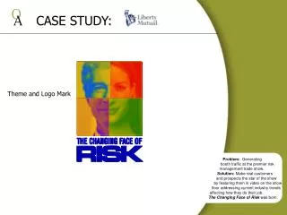
The view of myeloblasts on bone marrow aspiration material (×1000, Giemsa stain).
findings of bone marrow aspiration were correlated with AML-M2 with maturation type.
AML is a hematological malignancy characterized by proliferation of myeloid cell precursors with or without maturation. The initial symptoms of AML are related to anemia, neutropenia, and thrombocytopenia which develop due to bone marrow infiltration of leukemic blasts
Acute myeloid leukemia (AML) comprises 15–20% of all childhood acute leukemias. In most of the patients, fever, pallor, weight loss, and mucosal bleeding are seen. In more than half of the cases, liver, spleen, and lymph nodes are palpable
In FAB classification, AML is classified according to myeloblasts’ morphology into 10 subtypes I. n 2008, WHO classification was developed and AML was reclassified according to accompanying cytogenetic abnormalities. AML-M2 comprises 10–15% of all AML cases.