Download
1 / 60
600 likes | 735 Views
Unit 3. Cell Structure and Function. As Organisms Get Larger, Why do They Become Multi-cellular?. It’s all about the surface area to volume ratio!. Prokaryotic cells: Archaebacteria Eubacteria genetic material not in a nucleus no membrane bound organelles. Eukaryotic cells:

E N D
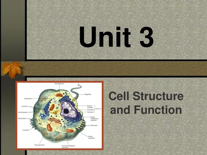
Unit 3 Cell Structure and Function
Prokaryotic cells: Archaebacteria Eubacteria genetic material not in a nucleus no membrane bound organelles Eukaryotic cells: Protists, Plants, Fungi and Animals true nucleus with genetic material has membrane bound organelles I. Prokaryotic vs. Eukaryotic Cells
A. Membranous organelles 1. Nucleus 2. Endoplasmic reticulum 3. Golgi apparatus 4. Mitochondrion 5. Chloroplast 6. Lysosomes 7. Peroxisomes II. Eukaryotic Cells B. Nonmembranous organelles 1. Ribosomes 2. Microtubules 3. Centrioles 4. Flagella 5. Cytoskeleton
Also called cytomembrane system part of the compartmentalization of the cell A. Endoplasmic reticulum a system of tubules and sacs continuous with the outer membrane of the nuclear envelope membranes are folded into sacs,cisternae,anddivide the cell into cytosol and cisternal space (lumen) 1. Smooth ER (no ribosomes) a. Location of synthesis of lipids (steroids, fats, phospholipids) b. Forms detoxification compartments (full of enzymes) for drugs and poisons c. Metabolism of carbohydrates III. Endomembrane system (eukaryotic)
2. Rough ER (ribosomes attached) location of the signal mechanism, i.e. how secreted proteins get into the cisternal space Membrane factory Proteins to be secreted from the cell have a signal sequence at the lead end (20-24 a.a.). When the signal sequence is produced in protein synthesis, a signal recognition particle (SRP) attaches to it and to the ribosome producing it. The SRP attaches the ribosome to a receptor protein on surface of ER. (hence “rough ER”) The signal sequence and subsequently the rest of the protein are transported (pushed) through the membrane into the cisternal space (lumen). 5-6) Signal sequence is removed, leaving the finished product (protein).
You have to read this!!!! Aoccdrnig to rscheearch at Cmabrigde Uinervtisy, it deosn't mttaer in waht oredr the ltteers in a wrod are, the olny iprmoetnt tihng is that the frist and lsat ltteer be at the rghit pclae. The rset can be a total mses and you can sitll raed it wouthit porbelm. Tihs is bcuseae the huamn mnid deos not raed ervey lteter by istlef, butthe wrod as a wlohe.amzanig huh?
3. Golgi apparatus (golgi body) stacks of sacs (cisternae), each stack is called a “dictyosome” How does Golgi apparatus work? a. ER containing protein pinches off to form a transport vesicle. b. Transport vesicle carries protein to internal surface of Golgi apparatus. c. A series of transport vesicles carries protein to outer cisternae from one cisterna to the next. d. In transit the proteins are processed, sorted, and modified. e. In cells that secrete proteins, the outer cisterna pinches off a secretory vesicle, which travels to the plasma membrane.
4. Lysosomes a. Formed as vesicles that bud off of the Golgi apparatus b. Filled with hydrolytic enzymes, most of which are effective at pH 5 (Why?) c. Some vesicles join food vacuoles = phagocytosis d. Some engulf and digest cell organelles = autophagy Tay Sachs is considered a storage disease because of a problem in the lysosome.
5. Peroxisomes Membrane-bound chambers where H+ is removed from various molecules and transferred to O2 to form H2O2 (hydrogen peroxide). Helps w/ breakdown of fatty acids and detoxifying poisons. c. Also contain enzyme catalase, which breaks down H2O2 into water because it is toxic itself
Hallmark Cards that should have been: • How could two people as beautiful as you… • …Have such an ugly baby? Congratulations Anyway! • I’ve always wanted to have someone to hold, someone to love. After having met you… • …I’ve changed my mind.
A. Microtubules 1. Structure a. Hollow, rod-shaped, 25 nm. in diameter b. Composed ofa and b tubulin dimers (globular proteins) IV. Cytoskeleton 2. Functions a. Structural • i. Radiate from the microtubule organizing center (MTOC), which is the centrosome • ii. Act as girders or as bundles near cell membrane, cell shape • iii. Very dynamic (form and reform) [Centrioles found in centrosome of animal cells are composed of 9 sets of 3 microtubules.]
b. Movement i. Organelles and vesicles move along microtubules (little train tracks), being pulled by a protein called kinesin. ii. Cilia and flagella, composed of “9 + 2” arrangement, use proteins called dynein as contractile side arms
iii. Basal bodies are found at the base of both cilia and flagella
B. Microfilaments helix of actin molecules (globular proteins), 7nm. 1. Structural - in microvilli, cell shape, dynamic 2. Functional - involved in cytokinesis, pseudopodia, and muscle contraction C. Intermediate filaments • heterogeneous, fibrous proteins, 8-12 nm. 1. Structural - Cell shape and stability, dynamic
- all cells have them - complex, dynamic structures, not passive - differentially permeable V. Plasma Membrane
A. Functions 1. Regulates the movement of molecules into and out of the cell 2. Site of cell recognition and communication B. Structure (Fluid mosaic model) (10nm.) 1. Phospholipids a. 5 - 10 different types b. Most common = phosphotidylcholine c. Membrane fusion easily accomplished d. Movement - 2 mm./sec. 2. Cholesterol - decreases fluidity of membrane
Membrane Fluidity • Why is it that membrane phospholipids drift laterally, and rarely flip?
How is this fluidity maintained? • Kinks in unsaturated fatty acid tails of phospholipids. • Cholesterol
3. Proteins Integral proteins -part or all the way through membrane (some move like icebergs) b. Peripheral proteins - usually bound to integral proteins (inside surface) 4. Glycoproteins and glycolipids - cell recognition - external surface oligosaccharides (<15 units)
C. Transport across the membrane 1. Diffusion movement of molecules down the concentration gradient passive process - energy from kinetic energy of molecules a. Channel proteins - aqueous channel for ions, gated
b. Facilitated diffusion - molecule binds to protein and is transported across membrane i. Protein changes its shape ii. Specificity
c. Osmosis - diffusion of water through a selectively permeable membrane
Know and understand these terms = isotonic, hypertonic, hypotonic, water potential, turgor, plasmolysis
Lab 1E - Plasmolysis #1 Onion Cells - Sketch Homework: Complete Analysis ?’s 1-3 on pg. 18 (Due Tom.) • Plasmolysis video clip • Animation
2. Active transport a. Movement of molecules up the concentration gradient b. Requires energy (ATP) c. E.g. sodium-potassium pump