Download
1 / 61
610 likes | 767 Views
Introduction to different brain and other clinical imaging methods. Oury Monchi, Ph.D. Parkinson Cognition Action & Neuroimaging (PCAN) Laboratory Centre de Recherche , Institut Universitaire de Gériatrie de Montréal & Universit é de Montréal http:// unfweb.criugm.qc.ca/oury.

E N D
Introduction to differentbrain and otherclinicalimagingmethods Oury Monchi, Ph.D. Parkinson Cognition Action & Neuroimaging (PCAN) Laboratory Centre de Recherche, InstitutUniversitaire de Gériatrie de Montréal & Université de Montréal http://unfweb.criugm.qc.ca/oury
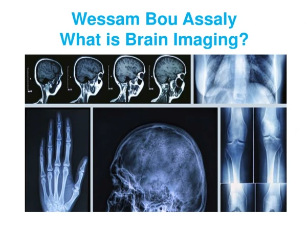
Imaging Techniques • MagneticResonance Imaging (MRI) • Brainanatomy techniques (volumetry, DTI) • Brainfunction technique (fMRI) • Vascular and heartimaging • Positron Emission Tomography (PET) • Single Photon Emission ComputedTomography (SPECT) • Application to Exercise Sciences
MRI Basic Principles • Magnet: • Very powerful (1 to 7T) and homogeneous and static magnetic field, it incites the hydrogen protons to align themselves. • Earth magnetic field 0.00005T! • Radiofrequency coils: • Generates and receive transient electromagnetic field, at the frequency of resonance of hydrogen disrupting alignment of protons from low to high energy state. • Energy released can be detected as they return to their base state • Speed to return to base states depends of the tissue they are part of, this generate the T1 and T2 signals. • Gradient coils: • Gradual fields aligned in x, y, z axes • Allows us to place detected signals in a 3D volume
Morphological variations • Large variabilityfrom one brain to the other • Can wemakeinferencesbased on population criteria (age, sex, health) on thismorphological basis? Whatcriteria do we use?
Techniques • Volumetry • VoxelBasedMorphometry (VBM) • Diffusion Tensor Imaging (DTI)
Principles of volumetry • An anatomical image allows us to separate the grey and white matter • One can paint the region of interest on each subject’s scan • We can study the variation of this region compared with a specific parameter (age, neuropsychological score, etc) or different groups (Parkinson’s vs healthy controls)
Volumetry: example • Womensufferingfromsomatoformdisorderscompared to control participants • Significantdifference in caudate nucleus volume Hakalaet al. (2004)
VoxelBasedMorphometry:principles • VBM consists in comparing local grey matter density between two populations • This comparison is not dependent on: • any particular structure • the experimenter’s subjectivity (as in volumetry, where regions are painted manually) • VBM is performed on the entire brain
VoxelBasedMorphometry:methods • Normalization to a template • Segmentation • Spatial smoothing • Statisticalanalysis
VoxelBasedMorphometry:applications • 22 Controls and 56 MCI (13 have evolvedintodementia) are followed over 22 months • Comparedwith stable MCIs, progressive MCIsexhibitatrophy in differentregions Hamalainenet al. (2007)
Diffusion Tensor Imaging:principles • Allows to obtain images based on properties of water molecule displacement in tissues • Reflects tissue properties (position, orientation, anisotropy), especially of white matter • Reflects tissue degradation (axons, myelin, cell wall) • Made possible by an adequate acquisition sequence
Concept of Diffusion:isotropy and anisotropy • Diffusion is isotropic if it is with the same amplitude in all directions • Diffusion is anisotropic if it prefers one or more directions • Fractional anisotropy characterizes local diffusion (1 > FA > 0)
Concept of MeanDiffusivity • Isotropyis not enough to characterize diffusion:
DTI: MRI sequence • One or more images at b=0 (T2 contrast) • As many image acquisitions as there are directions at b ~ 1000 sec/mm2
DTI: mapsobtained • Meandiffusivitymap • high signal in ventricles and sulci • Fractionalanisotropymap
DTI: FA and MD applications • Influence of age on meandiffusivity in grey and white matter • Correlation of bothmeasureswithage in greymatter, only in peakheight in white matter • Fibre reconstruction: average of 86 000km in aged participants comparedwith 118 000km in young Benedetti et al. (2006)
a g b c f e DTI: Fibertracking Study of anatomicalconnectivity
Basic Principles of fMRI • For a long time, a relationshipbetweenbrainactivity and deoxygenatedhemoglobin (whichisparamagnetic) in the blood has been known • In the early 90's itwasdiscoveredthat an MR pulse sequencecouldmeasure the rate of deoxygenatedhemoglobin (Thulborn et al.; Ogawa et al.) • This gave rise to Blood OxygenationLevelDependent (BOLD) fMRI or T2* sequence, whichprovides us with an indirect measure of brainactivity.
Functional MRI: Voice recognition Belin, et al. (2000) Nature
MR-measurement of aorticcompliance • Compliance of aortaishighlypredictive of overallvascularhealth • Flow velocityimagingallowsmeasurement of pressure-wave propagation in aorta • Vesselwallimagingallowsmeasurement of distensibility
Positron Emission Tomographyprinciples • PET depends on the injection of a radioactive isotope produced by a cyclotron • From the time of their injection, these radio-isotopes decay and emit positrons, whichcollidewithelectrons. These collisions produceoppositγ-raysthat are captured the coincidence detectors of the PET scanner • Depending on the moleculesthatthese isotopes bindwith, wecanget information on metabolism, blood flow, or the release of neurotransmitter (eg. 11C raclopridewhichbinds to striatal D2 receptors)
Radioactive tracers for PET • 18FDG (Fludeoxyglucose): glucose metabolism • H215O : regional blood flow (cerebral or myocardial) • 18FDOPA : Dopa uptake (dopamine precurser) • [11C]raclopride : Dopamine D2 antagonist • 18FP-TZTP : muscarinic agonist (acetylcholine) • PHNO, FLB 457, WAY, ……….
FDG PET • FDG-PET scan in a boy with left parietal-temporal epilepsy showing decreased glucose metabolism in the left parietal and temporal lobes
Water PET • Regional cerebral blood flow (rCBF) is related to glucose and oxygen consumption. • Very sensitive to acute changes… • E.g., patients with Parkinson’s disease who received DBS on STN perform a joystick task while OFF- or ON-DBS. • Similar task-induced rCBF changes in the M1 in both condition, but greater changes in SMA. • Normalizing effect of DBS.