Download
1 / 34
421 likes | 960 Views
Updates on the evaluation and management of Azoospermia. Presented by Omar Chahal MD. Supervised by Dr Khaled El Sayyid. Definition of Infertility. Inability to conceive after 1 year of unprotected sexual intercourse Affects approximately 15% of couples 40% of cases involve a male

E N D
Updates on the evaluation and management of Azoospermia Presented by Omar Chahal MD. Supervised by Dr Khaled El Sayyid
Definition of Infertility • Inability to conceive after 1 year of unprotected sexual intercourse • Affects approximately 15% of couples • 40% of cases involve a male • 40% of cases involve a female • 20 % involve both sexes
FSH & LH • In the testis, LH stimulates steroidogenesis within Leydig cells by inducing the mitochondrial conversion of cholesterol to pregnenolone and testosterone • FSH binds to sertoli cells and spermatogonial membranes within the testis and is the major stimulator of seminiferous tubule growth during development & is essential for the initiation of spermatogenesis at puberty
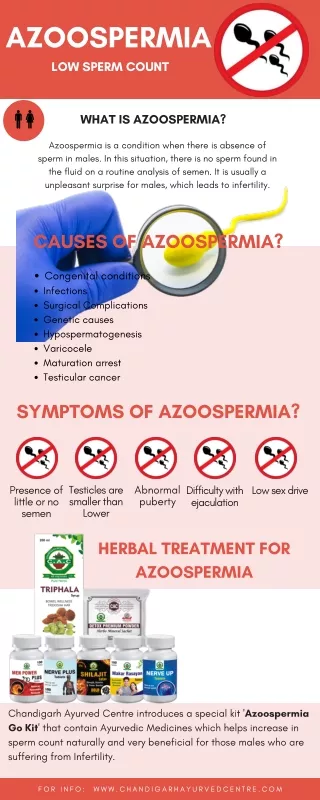
AZOOSPERMIA • Azoospermia is defined as complete absence of sperm from the ejaculate for at least two separate centrifuged semen samples • It is present in about 1% of all men and in approximately 15% of infertile men • Azoospermia is different from aspermia, in that aspermia is the complete absence of seminal fluid emission upon ejaculation
Causes of Azoospermia 1)Pretesticular:also called secondary testicular failure, usually endocrine in nature 2)Testicular:boardly termed as primary testicular failure, these are intrinsic disorders of spermatogenesis 3)Posttesticular:includes Ejaculatory dysfunction or obstruction of the genital tract, it constitute 40% of cases of azoospermia • Pretesticular and post-testicular causes are often amenable to treatment, which may restore fertility, whereas the success rates for intervention in testicular pathology are much more modest
Pretesticular • Hypothalamic disease * Gonadotropin deficiency (Kallmann syndrome) * Isolated LH deficiency * Isolated FSH deficiency * Congenital hypogonadotropic syndromes • Pituitary disease * Pituitary insufficiency (tumors, infiltrative processes, operation, radiation, deposits) * Hyperprolactinemia * Exogenous hormones (estrogen-androgen excess, glucocorticoid excess, hyper and hypothyroidism) *Growth hormone deficiency (prader-willi syndrome)
Testicular • Chromosomal (Klinefelter syndrome [XXY], XX sex reversal, XYY syndrome) • Noonan syndrome (male Turner syndrome) • Myotonic dystrophy • Vanishing testis syndrome (bilateral anorchia) • Sertoli-cell-only syndrome (germ cell aplasia) • Y chromosome microdeletions (DAZ gene) • Gonadotoxins (radiation, drugs) • Systemic disease (renal failure, liver failure, sickle cell anemia) • Defective androgen activity • Testis injury (orchitis, torsion, trauma) • Varicocele-induced testicular damage • Idiopathic
Posttesticular 1- Reproductive tract obstruction a. Congenital blockages: Congenital absence of the vas deferens (CAVD), example: Cystic Fibrosis Young syndrome (triad of chronic sinusitis, bronchiectasis, and obstructive azoospermia) Idiopathic epididymal obstruction PKD (secondary to obstructing cysts in the epididymis or seminal vesicle) Ejaculatory duct obstruction b. Acquired blockages: Vasectomy (performed for contraception) Groin surgery (iatrogenic injury of inguinal vas from hernia repair) Infection (which may involve the epididymis, with scarring and Obstruction) c. Functional blockages: Sympathetic nerve injury or medications that impair the contractility of seminal vesicle or Vasal musculature
Posttesticular 2- Disorders of sperm function or motility • Immotile cilia syndromes (Men have nonmotile but viable sperm in normal numbers) • Maturation defects (due to epididymal dysfunction after vasectomy induced Blockage) • Immunollogic infertility (may result from an abnormal exposure to sperm antigens after testicular injury)
Although both primary and secondary testicular failure will be associated with marked reduction in testicular volume, these entities can be distinguished by serum endocrine testing to include FSH, LH, testosterone, and prolactin levels • High serum FSH levels, typically greater than two times normal, are indicative of primary testicular failure, and diagnostic testicular biopsy is not required to rule out obstructive etiologies • Primary testicular failure in conjunction with azoospermia, commonly termed nonobstructiveazoospermia(NOA), is best managed with testicular sperm harvest for eventual ICSI • Azoospermic patients with normal testicular size, palpable vas deferens, and normal serum FSH levels require a diagnostic testicular biopsy to differentiate genital tract obstruction from disorders of spermatogenesis such as maturation arrest
Evaluation of specific conditions associated with azoospermia Ι. Absence of the vasadeferentia (vasal agenesis): The most common cause of congenital bilateral absence of the vas deferens (CBAVD) is a mutation of the cystic fibrosis transmembrane conductance regulator (CFTR) gene About 95% of males with clinical cystic fibrosis have CBAVD, and approximately 70% of men with CBAVD and no clinical evidence of a cystic fibrosis have an identifiable abnormality of CFTR gene The diagnosis of vasal agenesis, either bilateral or unilateral, is made by physical examination Imaging studies and surgical exploration are not necessary to confirm the diagnosis, but may be useful for diagnosing abnormalities associated with vasal agenesis
Due to the embryological association between the vasa and seminal vesicles, most patients with vasal agenesis also have seminal vesicle hypoplasia or agenesis. Since the majority of semen is derived from the seminal vesicles, almost all patients with CBAVD have low semen volume • In the azoospermic patient who has unilateral vasal agenesis, radiologic imaging with transrectalultrasonography (TRUS) may be useful to evaluate the ampullary portion of the contralateral vas deferens and the seminal vesicles, because unilateral vasal agenesis can be associated with contralateral segmental atresia of the vas deferens or seminal vesicle, resulting in obstructive azoospermia.
An abdominal ultrasound or CT scan should be obtained to assess anomalies such as renal agenesis • In 1 study, 26% of men with unilateral CAVD and 11% with CBAVD had Renal agenesis. This association between unilateral vasal agenesis and ipsilateral renal anomalies is due to their common embryological origin • However, since a man with CBAVD most likely has mutations in the CFTR gene, the Guidelines recommend genetic testing for CFTR mutations in the female partner before using sperm of a man with CBAVD
Π. Ductal Obstruction: a) Patients with normal ejaculate volume The serum FSH of a patient with normal semen volume is a critical factor in determining whether a diagnostic testicular biopsy is needed to establish the presence or absence of normal spermatogenesis In fact, FSH values in the upper normal range usually indicate impaired spermatogenesis while marked elevation of serum FSH is diagnostic of abnormal spermatogenesis – usually nonobstructiveazoospermia Although a diagnostic testicular biopsy will determine if spermatogenesis is impaired, it does not provide accurate prognostic information as to whether or not sperm will be found on future sperm retrieval attempts for patients with nonobstructiveazoospermia In addition, in cases of nonobstructiveazoospermia, the presence or absence of sperm in a diagnostic testicular biopsy specimen does not absolutely predict whether sperm are present elsewhere in that or the opposite testis
Therefore, a testicular biopsy is not necessary to either establish the diagnosis or to gain clinically useful prognostic information for patients with clinical findings consistent with the diagnosis of nonobstructiveazoospermia (i.e. testicular atrophy or markedly elevated FSH) Conversely, patients who have a normal serum FSH should undergo a diagnostic testicular biopsy The recommendation of the Male Infertility of AUA is to use the diagnostic testicular biopsy to distinguish between obstructive and non-obstructive causes of Azoospermia in patients with normal testicular size, at least 1 palpable vas deferens and a normal serum FSH level
If the testicular biopsy is normal, obstruction at some level in the reproductive system must be present and the location of the obstruction may then be determined Most men with obstructive azoospermia, palpable vasa and no history suggesting iatrogenic vasal injury have bilateral epididymal obstruction Epididymal obstruction can be identified only by surgical exploration
Testicular biopsy can be performed by a standard open incision technique or by percutaneous methods • A routine open testicular biopsy, performed under local anesthesia, is the most common method • This should be performed through a small scrotal incision without delivering the testis outside the skin or tunica vaginalis • This minimizes postoperative scarring and therefore facilitates subsequent scrotal reconstructive surgery
Vasography may be utilized to determine whether there is an obstruction in the vas deferens or ejaculatory ducts • The use of this technique is highly selective and should only be performed by urologists with microsurgical experience, who can proceed with reconstruction in the same setting, as obstruction can develop at the site of vasography
b) Patients with low ejaculate volume Low ejaculate volume (< 1.0 ml) that is not caused by hypogonadism or CBAVD can be caused by ejaculatory dysfunction, but is most likely caused by ejaculatory duct obstruction (EDO) Ejaculatory dysfunction rarely, if ever, causes low ejaculate volume with azoospermia, although it is a well-known cause of aspermia or low ejaculate volume with oligospermia Additional seminal parameters that can be helpful in determining the presence of EDO are seminal pH and fructose, since the seminal vesicle secretions are alkaline and contain fructose However, the results of semen pH and fructose testing may be misleading when these tests are not properly performed and, therefore, many experts tend to give less weight to these parameters over other clinical findings
grossly dilated left seminal vesicle with wide intravesicular spaces Transrectalultrasonography (TRUS) is indicated for the diagnosis of EDO in men with low ejaculate volume and palpable vasa While vasography is an alternative diagnostic test for EDO, TRUS is minimally invasive and avoids the risk of vasal injury associated with vasography
The finding of midline cysts, dilated ejaculatory ducts and/or dilated seminal vesicles (greater than 1.5 cm in anteroposterior diameter) on TRUS is suggestive, but not diagnostic, of ejaculatory duct obstruction • Conversely, normal seminal vesicle size does not completely rule out the possibility of obstruction
Therefore, seminal vesicle aspiration (SVA) and seminal vesiculography may be performed under TRUS guidance to make a more definitive diagnosis of EDO • The presence of large numbers of sperm in the seminal vesicle of an azoospermic patient is highly suggestive of EDO
Recommendation : Transrectalultrasonography with or without seminal vesicle aspiration and seminal vesiculography, should be considered as an initial minimally invasive diagnostic choice to identify ejaculatory duct obstruction in azoospermic men with low ejaculate volume and bilateral palpable vasa In patients with ejaculatory duct obstruction demonstrated by TRUS, testis biopsy may be considered if needed to confirm normal spermatogenesis Vasography with or without testicular biopsy should be considered a second line choice to identify the site of reproductive tract obstruction in these patients, and should not be done unless reconstructive surgery is undertaken at the same surgical procedure
Surgical management • TURED is the standard treatment of ED obstruction • In a series of 46 men with low semen volume azoospermia or severe oligospermia, Schroeder-printzen et al used TURED to treat, 46% had improvement in semen quality and 20% initiated a pregnancy. A concern of this study is that 4% of patients who had sperm in the preoperative ejaculation specimen became azoospermic after treatment • Futhermore, 20% of treated patients experienced complications. Including watery, high volume ejaculate, prolonged cathetherization for gross hematuria, UTI, chronic epididymitis with recurrent pain, post-void dribbling and premature ejaculation
Although the success rate is not impressive and complications are not negligible, TURED may allow select patients to undergo assisted reproductive techniques with ejaculated sperm Given the potential complications of urinary sphincter injury, rectal injury and reflux of urine into the reproductive tract with the use of TURED An alternative treatment for ED obstruction was proposed. In 1995 jarow and zagoria published a report on the use of antergrade balloon dilatation of the ED with the patient under general anesthesia to recanalize an obstructed ED The authors preferred the antegrade approach as it is often difficult to catheterize the orifice of the ED transurethrally, especially with distortion of the anatomy that may be present with obstruction
Microsurgical reconstruction of the reproductive tract (vasovasostomy & vasoepididymostomy) It is successful in patients with obstructive azoospermia Following vasectomy reversal, for example, return of sperm to the ejaculate occurs in 70-95% of patients, and pregnancies are obtained without the need for assisted reproduction in 30-75% of couples
A very important factor influencing the likelihood of sperm returning to the semen and of pregnancy after vasectomy reversal is: • The number of years between vasectomy and attempted reconstruction • The length of the obstructive interval • the presence or absence of sperm in the intraoperative vas fluid; the gross appearance and quality of the sperm in the vas fluid • The length of the vas segment between the epididymis and the vasectomy site • The presence or absence of a sperm granuloma at the vasectomy site • The likelihood of pregnancy after vasectomy reversal is also heavily influenced by the age of the female partner
Intra Cytoplasmic Sperm Injection: The sperm requirement for egg fertilization has dropped from hundreds of thousands for IVF to 1 viable sperm for ICSI This has led to the development of aggressive new surgical techniques to provide sperm for egg fertilization from men with apparent azoospermia Clinical pregnancy rates reported in the recent literature range from 26-57% and delivery rates range from 18- 75%