Download
1 / 75
940 likes | 1.64k Views
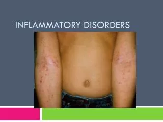
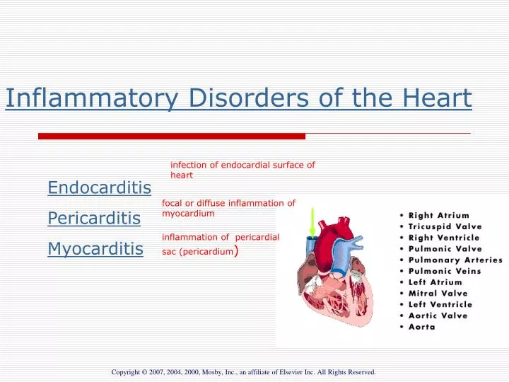
Inflammatory Disorders of the Heart. infection of endocardial surface of heart. Endocarditis Pericarditis Myocarditis. focal or diffuse inflammation of myocardium. inflammation of pericardial sac (pericardium ).

E N D
Inflammatory Disorders of the Heart infection of endocardial surface of heart Endocarditis Pericarditis Myocarditis focal or diffuse inflammation of myocardium inflammation of pericardial sac (pericardium)
Endocarditis: precipitated by bacteria/fungal infection; potential death from emboli and valvular disturbance Myocarditis: virus, toxin or autoimmune response damage heart muscle > lead to cardiomyopathy and death! Pericarditis: Bacterial, fungal or viral infection affect visceral and parietal pericardium; restrict heart pumping action> lead to cardiac tamponade and death!
Layers of the Heart Layers of heart muscle and pericardium; section of heart wall shows fibrous pericardium, parietal and visceral layers of serous pericardium (with pericardial sac between them), myocardium, and endocardium- Fig. 37-1
Infective Endocarditis(Click to access YOUTube video) • Infection of inner layer of heart- usually affects cardiac valves • Was almost always fatal until development of penicillin • 15,000 cases diagnosed in US each year
A- Aortic Valve B- Mitral Valve C- Tricuspid Valve - Pulmonary Valve
A&P Review- Blood enters right atrium and moves through _______ into right ventricle. Blood then moves from right ventricle into pulmonary artery via _________. • After entering left atrium via pulmonary veins, blood moves through the _____ into left ventricle. • Finally, it travels through the _____ and out of heart A- Aortic Valve B- Mitral Valve C- Pulmonary Valve D- Tricuspid Valve A- Aortic Valve B- Mitral Valve C- Pulmonary Valve D- Tricuspid Valve
Risk Factors- endocarditis • Hx of rheumatic fever or damaged heart valve • Prior history of endocarditis • Invasive procedures- (introduce bacteria into blood stream) dental,gyne, etc. • Recent Dental Surgery • Permanent Central Venous Access • IV drug users • Valve replacements
Classification • Subacute form (subacute bacterial endocarditis-SBE) • Gradual onset; longer clinical course • Caused by enterococci • Usually those with damaged valves • Acute form • Shorter clinical course • Abrupt onset • Usually those with healthy valves • Usually caused by staph aureus • *Classify by cause as IVBA; prosthetic valve endocarditis (PVE), fungal endocarditis
Causative Organisms • Most common causative organism • Streptococcus viridans • Staphylococcus aureus • Viruses • Fungi
Etiology and Pathophysiology • Key -Blood turbulence within heart allows causative agent to infect previously damaged valves or other endothelial surfaces • Principal risk factors • Prior endocarditis • Prosthetic valves • Acquired valvular disease • Cardiac lesions
When valve damaged, blood > slowed down > forms clot. • Bacteria > into blood stream • Bacterial or fungal vegetative growths deposit on normal or abnormal heart valves • Infection of innermost layers of heart may occur in people with: • congenital and valvular heart disease • history of rheumatic heart disease • normal valves with increased amounts of bacteria
Bacterial Endocarditis of Mitral Valve Bacterial endocarditis of mitral valve. Valve is covered with large, irregular vegetations (note arrow). From text
Etiology and Pathophysiology • Vegetation • Fibrin, leukocytes, platelets, and microbes • Adhere to valve or endocardium • Embolization of portions of vegetation into circulation
Sequence of Events in Infective Endocarditis (view carefully) Fig. 37-3
Clinical Manifestations • Nonspecific • Fever occurs in 90% of patients • Chills • Weakness • Malaise • Fatigue • Anorexia
Clinical Manifestations • Subacute form • Arthralgias • Myalgias • Back pain • Abdominal discomfort • Weight loss • Headache • Clubbing of fingers
Clinical Manifestations • Vascular manifestations • Splinter hemorrhages in nail beds • Petechiae • Osler’s nodes on fingers or toes • Janeway’s lesions on palms or soles • Roth’s spots • *Murmur in most patients • Heart failure in up to 80% with aortic valve endocarditis • *Manifestations secondary to embolism
Sites of emboli due to infective endocarditis (AKA metastic infections)-site determined by location of original lesion
Osler’s nodes Janeway lesions Splinter hemorrhages Roth spots
Osler’s nodes- painful, red or purple pea-sized lesions on toes and fingertips • Splinter hemorrhages- black longitudinal streaks on nail beds • Janeway lesions-flat, painless, small, red spots on palms and soles • Roth spots- hemorrhagic retinal lesions
Diagnostic Studies • History • Recent dental, urologic, surgical, or gynecological procedures • Heart disease; onset *new heart murmur • Recent cardiac catheterization • Skin, respiratory, or urinary tract infection • Laboratory tests • Blood cultures (if temp above 101, typically do 2 sets) • WBC with differential • ESR, CRP • Echocardiography- TEE best- see vegetations • Chest x-ray 1) Vegetations on mitral valve 2) Vegetations on aortic Valce
Collaborative Care • Prophylactic treatment for patients having (see prevention) • Removal or drainage of infected tissue • Renal dialysis • Ventriculoatrial shunts • Antibiotic administration • Monitor antibiotic serum levels (peak & trough) • Subsequent blood cultures • Renal function monitored • BUN, Creatinine
Collaborative Care • Antibiotic therapy cont • IV for 2-8 weeks • *Maybe oral meds if not a good candidate for IV and can identify and treat the specific causative organism • Fungal and prosthetic valve endocarditis • Responds poorly to antibiotics • Valve replacement- adjunct procedure • Fever • Comfort with ASA, Ibuprofen etc
Collaborative Care • Surgical/Therapeutic/Nursing • Early valve replacement. • Complete bed rest –only if temp remains elevated or signs HF • Overall goals • normal or baseline cardiac function • performance of activities of daily living (ADLs) without fatigue • Antibiotic therapy cont
Nursing Diagnoses • Risk for Imbalanced Body Temperature-Hyperthermia • Risk for Ineffective Tissue Perfusion-emboli • Risk for decreased cardiac output • Ineffective Health Maintenance • Deficient knowledge
Complications • Emboli (50% incidence) • Right side- pulmonary emboli (esp. with IV drug abuse- Why??) • Left side-brain, spleen, heart, limbs,etc • CHF-check edema, rales, VS • Arrhythmias- A-fib • Death .
Collaborative Care • Priority Teaching • Signs and symptoms of life-threatening complications of IE, as cerebral emboli, HF etc. • Monitor fever (chronic or intermittent)- sign that drug therapy ineffective • Monitor lab data, blood cultures- determine effectiveness of antibiotic therapy • Critical-prophylactic antibiotic therapy prior to ANY invasive procedure (*changes in requirements- see later slide)
Collaborative Care • Priority Teaching/nursing care • Stress need to avoid infectious people • Avoidance of stress and fatigue • Manage rest, hygiene, nutrition • Assessment of nonspecific manifestations • Monitor laboratory data • Monitor patency of IV • Teach reduction measures for risk for infection • Stress follow-up care
Collaborative Care • Eliminate risk factors • Patient teaching • Penicillin prophylaxis • Recent change 2007 guidelines (not all require prophylaxis) • if have prosthetic valve • History of endocarditis • Certain congenital heart defects • Heart transplant recipients- at risk • Removal or drainage of infected tissue • Renal dialysis • Ventriculoatrial shunts • Prevention
Risk Stratisfication for IE High Risk- • Mechanical prosthetic heart valve • Natural prosthetic heart valve • Prior infective endocardititis • Valve repair with prosthetic material • Most congenital heart diseases Moderate Risk- • Valve repair without prosthetic material • Hypertrophic cardiomyopathy • Mitral valve prolapse with regurgitation • Acquired valvular dysfunction Low Risk- • Innocent heart murmurs • Mitral valve prolapse without regurgitation • Coronary artery disease • People with pacemakers/ defibrillators • Prophylactic antibiotics are generally recommended only for people in the “High Risk” category
Pericarditis(Click to access YouTube video) • Pericarditis • inflammation of pericardium, thin, fluid-filled sac surrounding heart. • Can cause severe chest pain especially upon taking a deep breath) • Shortness of breath; hear pericardial friction rub.
Etiology/Pathophysiology • Pericarditis due to • bacterial, fungal or viral infection • heart loses natural lubrication(10-30cc’s); layers roughen and rub • Inflammatory process causes lymphatic fluid build-up- • if sudden > cardiac tamponade- • Pericardial Effusion- usually 250 cc’s before show up on x-ray-Can have 1000cc’s.
Normal Pericardium A&P 1 Heart part 1 Click here for information on pericardium, pericarditis
Fig. 37-4 Hear friction rub Acute pericarditis. Note shaggy coat of fibers covering surface of heart.
Risk Factors/pericarditis • Be acute or chronic • Post MI (Dressler’s syndrome)-4-6 wks • Secondary to chemo and cancer • Secondary to uremia in renal failure-40-50% of pts will develop • Trauma or cardiac surgery • AIDS- has emerged as most frequent CV manifestation • If chronic disorder-pericardium becomes rigid
Clinical Manifestations • Inflammation and pain • Friction rub- *diaphragm at L L sternal border- client sitting, leaning forward, best heard during inspiration (Pericardial Friction Rub) • Fever • Substernal, sharp, pleuritic chest pain • Inc. with coughing, breathing,turning, lying flat • Dec. with sitting up and leaning forward • Referred to trapezius muscle • Dyspnea
Complications of Pericarditis • Pericardial Effusion • Cardiac Tamponade
Pericardial Effusion • Can occur rapidly or slowly • Pulmonary compression-cough, dyspnea, and tachypnea • Phrenic nerve involvement- hiccups • Laryngeal nerve- hoarseness • *Slow build-up; no immediate effects; rapid lead to compression of heart (tamponade)
Pericardial Effusion- EKGElectrical Alternans • Pericardial effusion with electrical alternans • The QRS axis alternates between beats. In this example it is best seen in the chest leads where the QRS points in different directions! • This is rarely seen and is due to the heart moving in the effusion.
Cardiac Tamponade • Compression of heart • Occur acutely (trauma) or sub-acutely (malignancy) • Symptoms- chest pain, confusion, anxious, ^ CVP, restless, muffled heart sounds • Later- tachypnea, tachycardia, and dec. CO, NVD and pulsus paradoxus • With slow onset dyspnea may be only symptom • If rapid compression-Medical Emergency
PERICARDIUM CARDIAC TAMPONADE Original heart size Excess pericardial fluid
Definition- a decrease in systolic BP with inspirations that is exaggerated in cardiac tamponade
Determination of Pulsus Paradoxus • Place patient in position of comfort; take systolic BPduring baseline respiration. • Raise sphygmomanometer pressure until Korotkoff sounds disappear. • Lower pressure slowly until first Korotkoff sounds are heard during early expiration with their disappearance during inspiration • Record this pressure. • Very slowly lower pressure (1mm at a time) until Korotkoff sounds heard throughout respiratory cycle with even intensity. • Record this pressure. • The difference between the two recorded pressures is Pulsus Paradox. • Hemodynamically significant pulsus paradox is greater than or equal to 10 but look at trends. People with COPD may have a paradox due to increased thoracic pressures.
Collaborative Care-Pericarditis, Pericardial Effusion, Cardiac Tamponde • Diagnostic Tests • Medications • Surgical/Therapeutic Interventions • Nursing Diagnosis/Interventions
Diagnostic Tests- to R/O • CBC-inc. WBC, ESR, and CRP • Cardiac Enzymes- inc. but not as much as with MI • EKG- St elevation, Twave inversion • Echo- for wall movement • CXR • CT or MRI- for pericardial effusion • Pericardiocentesis fluid- determine cause