Download
1 / 70
700 likes | 949 Views
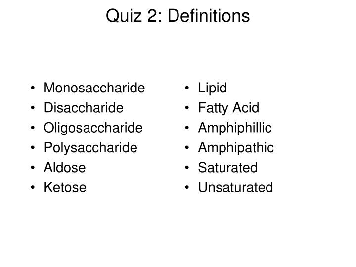
Quiz 2: Definitions. Monosaccharide Disaccharide Oligosaccharide Polysaccharide Aldose Ketose. Lipid Fatty Acid Amphiphillic Amphipathic Saturated Unsaturated. Quiz 2: Identification (Carbs). Quiz 2: Identification (Lipids). Protein Structure III. Relevent Interactions.

E N D
Quiz 2: Definitions • Monosaccharide • Disaccharide • Oligosaccharide • Polysaccharide • Aldose • Ketose • Lipid • Fatty Acid • Amphiphillic • Amphipathic • Saturated • Unsaturated
Relevent Interactions • Hydrophobic Forces • Electrostatic forces • Ion Pairs • Dipole–Dipole Interactions • Hydrogen Bonding • Covalent Bonds
Hydrophobic Forces(Entropic) Minimizes order of solvent H2O which occurs when hydrophobic molecules are in aqueous environment Primary Determinant of Tertiary and Quaternary Structures
Hydrophobic Interactions Not “bonds”; exclusion of water Buried in interior Important in tertiary structure
Electrostatic Forces • Ion Pairs • Attractive or Repulsive • Coulomb’s Law (strength proportional to 1/r2 • Competition between buried ionic interactions and hydrated ionic species on the surface
Ion Pairs Strength dependent on magnitude of charges, dielectric constant, and distance Modest strength Contribute little to native protein structure
Ion Pairs or Salt Bridges(Myoglobin) Figure 6-36
Ion-Polar Bonds Similar to electrostatic bonds Alternative to interactions with water Contribute little to native protein structure
Dipole-Dipole Interactions • Van der Waals Attractions • Strength proportional to 1/r6 • Dipole–Dipole Interactions • Dipole-Induced Dipole Interactions • Induced Dipole-Induced Dipole Interactions (London Dispersion Forces) Significant Contribution to Protein Native Structure
Typical Hydrogen Bonds between side chains(unshared electron pair: N and O)
Hydrogen Bonds Strength greatest in a polar environment Contribute greatly to secondary structure
Other Hydrogen Bonds(e.g. –SH)Similar electronegativity as –CH3More polarizable than –CH
Covalent Bonds Disulfide Bonds
Covalent (Disulfide) Bonds Do not need to be adjacent in primary structure Strong Intra- or Interchain
Sum of Forces Conformational Stability e.g. linking of fingers
Flexibility(conformational changes possible) Interaction (binding) of small molecules (effectors) Modification of protein amino acids – e.g. phosphorylation of serine
Cys2–His2 Zinc Finger Motif Stabilization of Small Domain Figure 6-37
Molecular Dynamics of Myoglobin Proteins Are Dynamic Structures Figure 6-38
Tertiary Structure Folding and ordering of a polypeptide chain due to interactions involving the amino acid side chains
Characteristics of Tertiary Structure • May contain both helices and sheets • Structural characteristics • Nonpolar residues: interior • Charged residues: surface (hydrated) • Polar residues: surface (hydrated) or interior (hydrogen-bonded) • Compact: little or no internal space for water molecules • Domain Structure
Side Chain Distribution in Horse Heart Cytochrome c Figure 6-27
Tertiary Structures Contain Combinations of Secondary Structure Motifs
Super Secondary Structural Motifs Greek Key hairpin Figure 6-28
Two Domain Protein(glyceraldehyde-3-P dehydrogenase) Figure 6-31
Quaternary Structure Specific association of polypeptide chains Subunits
Characteristics of Quaternary Structure • Identical or nonidentical subunits • Subunits usually associate noncovalently • Subunits are symmetrically arranged • Efficient means of producing highly complex proteins • Basis for regulatory behavior of many enzymes
Quaternary Structure of Hemoglobin (22) Figure 6-33
Three Broad Categories of Proteins • Fibrous Proteins • Globular Proteins • Membrane Proteins
Characteristics of Fibrous Proteins • Rod-like • Insoluble • due to hydrophobic AAs both inside and outside • Structural
-Keratin: Evolved for Strength (Hair, Wool, Nails) Right handed a-helix Left handed coiled coil
Coiled Coil non-polar residues Figure 6-15a
Collagen — A Triple Helical Cable • Component of connective tissue • Distinctive amino acid compostion • ~33% glycine • 15-30% 4-hydroxyproline • Some 3-hydroxyproline & 5-hydroxylysine • Right-handed triple helix • Organized into fibrils • Fibrils are covalently cross-linked • Collagen defects are responsible for a variety of human diseases
Collagen: in connective tissue, cartilage, gelatin • left-handed-helix • 3 AA per turn • 3 -chains are supertwisted • mainly Gly, Ala, Pro • (Gly-X-Y motif) • 6% hydroxy-proline confers thermostability
Proline Hydroxylase Requires ascorbic acid (vitamin C) Scurvy
Properties of Globular Proteins • Majority of proteins • Dynamic Functions, i.e. enzymes • Mixture of secondary structures • Soluble • hydrophobic core, polar surface
Membrane Proteins: receptors, transporters, enzymes, ion channels Hydrophobic Water (aqueous) Non-Polar Water (aqueous)
Determination of Tertiary Structure X-Ray Crystallography Nuclear Magnetic Resonance (NMR)
X-Ray Diffraction Pattern Figure 6-21