Download
1 / 37
380 likes | 522 Views
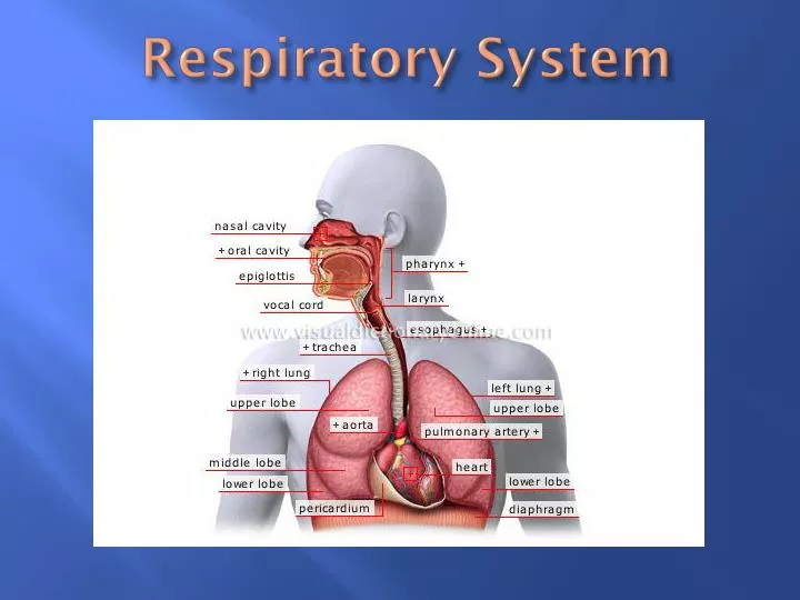
Respiratory System. Respiratory System – Fig. 14-1. Includes the passages that transport air to and from the lungs, and the air sacs in which gas exchange occurs. Respiration is the entire process by which gases are exchanged between the atmosphere and the body cells.

E N D
Respiratory System – Fig. 14-1 • Includes the passages that transport air to and from the lungs, and the air sacs in which gas exchange occurs. Respiration is the entire process by which gases are exchanged between the atmosphere and the body cells. • http://www.youtube.com/watch?v=o2OcGgJbiUk&feature=related (Overview of respiratory system) • (Don’t get confused with cellular respiration which is the breakdown of food to produce chemical energy.)
Organs or the Respiratory System • Upper respiratory (outside thorax) – nose, larynx, & pharynx • Lower respiratory (within thorax) – trachea, bronchial tree, and lungs • Nose • Supported by bone & cartilage • Nostrils provide entrances for air
Organs or the Respiratory System • Nasal Cavity (space behind the nose) – Fig. 14-4 • Divided medially by the nasal septum • Nasal conchae divide the cavity into passageways & help increase the surface area of the mucous membrane • Mucous membrane (Fig. 14-2) – filters, warms & moistens incoming air; particles trapped in the mucus are carried to the pharynx by ciliary action & are swallowed
Organs or the Respiratory System Cilia Pseudostratified epithelium Submucosa Mucous gland
Organs or the Respiratory System • Sinuses – Fig. 14-3 • Spaces in the bones of the skull that open into the nasal cavity • Lined with mucous membrane that is continuous with the lining of the nasal cavity • Function mainly to reduce weight of skull, they also serve as resonant chambers that affect the quality of the voice
Organs or the Respiratory System • Pharynx (throat) • Located behind the mouth and between the nasal cavity and the larynx • Functions as a common passage for air and food • Aids in creating vocal sounds
Organs or the Respiratory System • Larynx – Fig. 14-5 • Enlargement at the top of the trachea • Serves as passageway for air & helps prevent foreign objects from entering the trachea • Composed o muscles & cartilage • Contains the vocal cords which produce sounds by vibrating as air passes over them • Pitch of sound is related to the tension of cords • Intensity is related to the force of the air passing over the cords • Glottis (space between vocal cords) & epiglottis help prevent food & liquid from entering the trachea
1- vocal cords; 2 – vestibular fold; 3 – epiglottis; 4 – glottis; 5 – cartilage; 6 – sinus piriformis; 7 – base of the tongue
Organs or the Respiratory System • Larynx – Fig. 14-5 • http://www.youtube.com/watch?v=ajbcJiYhFKY (vocal cords)
Organs or the Respiratory System • Trachea – Fig. 14-6 • Extends into the thoracic cavity in front of the esophagus • Divides into the right and left bronchi • Mucous lining continues to filter incoming air • Wall is supported by incomplete (C shaped) cartilaginous rings; keeps trachea from collapsing
Organs or the Respiratory System • Bronchial tree – • Consists of branched air passages that lead from the trachea to the air sacs • Primary bronchi secondary bronchi bronchioles alveolar ducts alveolar sacs alveoli • ****Respiratory surface is alveoli – exchange of carbon dioxide and oxygen between alveoli & capillaries of circulatory system****
Organs or the Respiratory System • Bronchial tree (con’t) • Structure of the respiratory tubes • As tubes branch the amount of cartilage in the wall decreases and the muscular layer becomes more prominent • Elastic fibers in the walls aid the breathing mechanism • Epithelial lining changes from pseudostratified & ciliated to cuboidal & simple squamous as tubes decrease in size
Organs or the Respiratory System • Lungs • Left and right lungs are separated by the mediastinum and are enclosed by the diaphragm & the thoracic cage • Visceral pleura is attached to the surface of the lungs; parietal lines the thoracic cavity • R. lung – 3 lobes; L. lung – 2 lobes • Lobes contain – alveoli, nerves, blood vessel, and lymphatic & connective tissue (no muscle tissue)
Breathing/Pulmonary Ventiliation – Fig. 14-11 – movement of air into & out of lungs • Inspiration – air moves into lungs (atm. Pressure = 760 mm Hg) • Phrenic nerve impulses cause diaphragm to contract • As dome shaped diaphragm moves downward the size of the thoracic cavity increases • External intercostal muscles may contract causing ribs to rise which causes in increase in the size of thoracic cavity • As size of thoracic cavity increases pressure within alveoli DECREASES (758 mm Hg) • Atm. Pressure which is greater on the outside forces air into respiratory tract • Lungs inflate
Breathing/Pulmonary Ventilation – Fig. 14-11 – movement of air into & out of lungs • Expiration – air moves out of lungs • Diaphragm & external respiratory muscles relax • Elastic tissue of the lungs, thoracic cage and abdominal organs, which were stretched during inspiration suddenly recoil & surface tension causes alveolar walls to collapse • Pressure within the alveoli INCREASES (761 mm Hg) causing expiration of air out of lungs
Breathing/Pulmonary Ventilation – Fig. 14-11 – movement of air into & out of lungs • http://www.youtube.com/watch?v=q6-oyxnkZC0 (Boyles Law and ventilation)
Breathing/Pulmonary Ventilation – Fig. 14-11 – movement of air into & out of lungs • Respiratory or Pulmonary Air Volumes & Capacities – Fig. 14-13 • Tidal volume – amount of air that normally moves into or out of lungs during quiet breathing (approx. 500 ml) • Inspiratory reserve volume – additional air that can be inhaled (approx. 3000 ml) • Expiratory reserve volume – additional air that can be exhaled (approx. 1100 ml) • Residual volume – amount of air that remains in lungs (approx. 1200 ml)
Breathing/Pulmonary Ventilation – Fig. 14-11 – movement of air into & out of lungs • Respiratory or Pulmonary Air Volumes & Capacities – Fig. 14-13 • Vital capacity – maximum amount of air a person can exhale after taking the deepest breath possible • VC = TV + IRV + ERV • Lung Capacity – TLC = VC + RV – varies with age, sex, body size, athlete vs. non athlete • Residual air mixes with new air, this prevents concentrations of oxygen and carbon dioxide from fluctuating excessively with each new breath
Control of Breathing – normal breathing is rhythimic & involuntary, although the respiratory muscles can be controlled voluntarily • Respiratory Center • Located in the brain stem and includes parts of the medulla oblongata & pons • Controls both inspiration & expiration & rates of breathing
Control of Breathing • Factors affecting breathing • Breathing is affected by certain chemicals, the stretching of lung tissues & the persons emotional state • Chemosensitive areas are associated with the respiratory center • CO2 combines with H2O to form H2CO3 which in turn dissociates into HCO3- & H+ • Chemosensitive areas mainly influenced by [H+], this concentration determines acidity of blood • Stimulation of these areas causes the breathing rate to increase
Control of Breathing • Factors affecting breathing • Chemoreceptors in the carotid & aortic bodies • Sensitive to low [O2] • When [O2] is low increase breathing rate • An inflation reflex is triggered by stretching the lung tissues; this reflex reduces the duration of inspiratory movements & prevents over-inflation of the lungs during forceful breathing
Alveolar Gas Exchange – Internal Respiration • Alveoli • Tiny sacs clustered at the distal ends of the alveolar ducts • Some alveoli have openings into adjacent air sacs that provide alternate pathways for air when passages are obstructed • Respiratory Membrane/Surface • Consists of the alveolar & capillary walls • Gas exchange occurs here • Diffusion • Gasses diffuse from regions of higher concentration to lower concentration • O2 diffuses from alveoli to capillaries; • CO2 diffuses from capillaries to lungs
Transport of Gases – blood transports O2 & CO2 – Fig. 14-12 • O2transport • Mainly combines with HB (hemoglobin) to produce oxyhemoglobin O2 + Hb HbO2 • HbO relatively unstable & releases O2 in areas where [O2] pressure is low • There is an increase in the release of O2 from the blood when • CO2 levels increase • pH becomes more acidic • blood temperature increases
Transport of Gases – blood transports O2 & CO2 – Fig. 14-12 • CO2transport • Carried in solution either bound to Hb or as HCO3- • Most CO2 transported as HCO3- (approx. 70%) • CO2 + HbHbNHCOOH (carbamino hemoglobin) (approx. 23%) • Some dissolved in plasma (approx. 7%)
Transport of Gases – blood transports O2 & CO2 – Fig. 14-12 Carbon monoxide – CO • Combines with Hb more readily than O2 & forms a stable compound • Toxic because decreases Hb available for transport of O2 • Utilization of O2 – used for cellular respiration • C6H12O6 + 6O2 6CO2 + H2O + ATP