Download
1 / 69
710 likes | 922 Views
Mechanisms of Cell Invasion. Cancer Biology Course Feb 26, 2008. Outline. Background Material on Invasion & Metastasis Human tumor histopathology Culture Models Molecular Mechanisms Stromal / Tumor Cell Interactions Orthotopic Model for Skin Cancer Sahai paper Invadopodia Meeting.

E N D
Mechanisms of Cell Invasion • Cancer Biology Course • Feb 26, 2008
Outline • Background Material on Invasion & Metastasis • Human tumor histopathology • Culture Models • Molecular Mechanisms • Stromal / Tumor Cell Interactions • Orthotopic Model for Skin Cancer • Sahai paper • Invadopodia Meeting
Reading Material • Cell Biology Text - Molecular Cell Biology, Lodish 6th Edition: Chaps 17, 18 & 19. • Cancer Biology Text - The Biology of Cancer, Weinberg, 2007. Chap 14. • Original Article for Discussion • Gaggioli et al. 2007. Fibroblast-led collective invasion of carcinoma cells with differing roles for RhoGTPases in leading and following cells. Nat Cell Biol. 9:1392-1400.
Online Material • Bob Weinberg / MIT. “How Cancer Begins” - Streaming Podcast Lecture • http://mitworld.mit.edu/play/149/ • Cell Migration Consortium • Lists of reagents, background info, literature • http://www.cellmigration.org/resource/ • Invadopodia/Podosome Consortium - New • http://www.invadosomes.org/
Breast Cancer Slide Link Link - Click Here
Metastasis via Lymphatics Lymphatic Drainage of the Breast
Metastasis via Lymphatics Identification of “Sentinel” Lymph Node with Dye
Metastasis via Lymphatics H&E Staining: Breast Ca in Lymph Node
Metastasis via Lymphatics Keratin Staining: Breast Ca in Lymph Node
Culture Models of Invasion • Simple Criteria for Transformation • Loss of contact inhibition of growth • Growth in soft agar • Tumors in nude mice • Cells on Fluorescent Matrix • Cells in 3-D Matrix • Orthotopic Models - Sahai paper
Cells on Fluorescent Matrix Actin Matrix Smooth Muscle Cell Matrix Actin Macrophage
Growth Factor Pathways & Oncogenes • Receptor Tyr Kinases • EGFR, PDGFR, VEGFR • Cytoplasmic Tyr Kinases • Src, Syk-ZAP70, Abl • Small G-proteins • Ras
Connections from Integrins & Srcto Cell Migration • Integrins activate Src & FAK • Tyrosine kinases • Focal contacts / adhesions • Paxillin & Vinculin
Connections from Integrins & Srcto Cell Migration • Integrins activate Src & FAK • Tyrosine kinases • Focal contacts / adhesions • Paxillin & Vinculin • Cortactin - Src Substrate • Stabilizes Arp2/3 Networks • Signaling Scaffold
Connections from Ras-like Proteinsto Cell Migration • Rac - Lamellipodia • WAVE family of Arp2/3 activators • Cdc42 • WASp family of Arp2/3 activators • RhoA • Myosin-II Activation for Contraction • Others • Formins - mDia • CARMIL?
Tumor / Stroma:Paracrine Scenario a: Tumor cells activate surrounding stromal cells, i.e., fibroblasts, macrophages, and blood vessels. b: Fibroblasts respond by undergoing smooth muscle differentiation, blood vessels by angiogenesis, and both myofibroblasts and macrophages secrete additional cytoklnes. c: Modulated extracellular matrix production derives primarily from activated myofibroblasts, recruited vascular cells, and leaky blood vessels. d: Tumor cells, myofibroblasts, and activated macrophages cooperate in proteolytic action at invasive front. e: In the tumor center, angiogenesis is inhibited, tissue undergoes extensive fibrosis, and tumor cells may become necrotic.
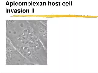
Sahai paper - Orthotopic model of cancer cell invasion • Gaggioli et al. 2007. Fibroblast-led collective invasion of carcinoma cells with differing roles for RhoGTPases in leading and following cells. Nat Cell Biol. 9:1392-1400.
Confocal Microscopy Pinhole Focal Plane
Two-Photon Microscopy Thin Region of Illumination Two Photons Absorbed
Cells Crawling on Collagen Fibers Time-lapsed movie of high metastatic CFP-labeled cells (white) and low metastatic GFP-labeled cells (green) crawling on collagen fibers (purple) in vivo.
Lamellipodia / Ruffles in situ Time-lapsed movie of a CFP labeled cell with a ruffling lamella on a field of GFP labeled cells in a living tumor.
Ameboid Movements of Cells High metastatic CFP-labeled cells (white) and low- metastatic GFP labeled cells (green) crawling on collagen fibers (purple).
Orthotopic Model for Skin Cancer Invasion • SCC12 - Squamous cell carcinoma cells • Stromal fibroblasts - from cancers • Matrix - dense fibrillar collagen w/ laminin & collagen IV
Epidermal Cancer Cells Figure 1. Fibroblasts promote and lead collective SCC invasion. (a) H&E-stained sections of SCC12 cells cultured in an organotypic system in the absence or presence of stromal fibroblasts. Mesench Cancer Cells Untransf Keratinocytes
Movie 1 SCC cells invade as collective strands. Sequential confocal sections of SCC12 and A431 cells invading in co-cultures with tumour fibroblasts (stained with phalloidin): note how all cells are incorporated into invading chains.
Fibroblasts lead SCC cells (Upside-down images) Figure 1. Fibroblasts promote and lead collective SCC invasion. (f) Representative images of SCC12 cells (green) and stromal fibroblasts (red) invading into naïve matrix.