Download
1 / 33
330 likes | 535 Views
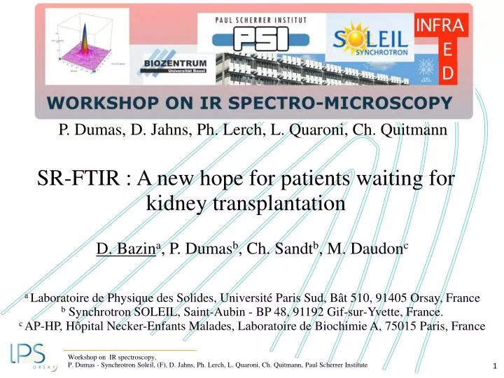
P. Dumas, D. Jahns, Ph. Lerch, L. Quaroni, Ch. Quitmann. SR-FTIR : A new hope for patients waiting for kidney transplantation. D. Bazin a , P. Dumas b , Ch. Sandt b , M. Daudon c a Laboratoire de Physique des Solides, Université Paris Sud, Bât 510, 91405 Orsay, France

E N D
P. Dumas, D. Jahns, Ph. Lerch, L. Quaroni, Ch. Quitmann SR-FTIR : A new hope for patients waiting for kidney transplantation D. Bazina, P. Dumasb, Ch. Sandtb, M. Daudonc a Laboratoire de Physique des Solides, Université Paris Sud, Bât 510, 91405 Orsay, France b Synchrotron SOLEIL, Saint-Aubin - BP 48, 91192 Gif-sur-Yvette, France. c AP-HP, Hôpital Necker-Enfants Malades, Laboratoire de Biochimie A, 75015 Paris, France
LectureI/ Generalities on pathological calcificationsII/ LithiasisIII/ Chemical diversity of intratissular renal calcificationsIV/ When intratissular calcification leads to ESRFA school case : DHADV/ Conclusion & Perspectives
(B2) From the medical point of view, pathological calcifications refer to either a concretion, e.g. a kidney stone [A], or an ectopic calcification [B] often associated with tissue alteration. Additionally, normal physiological calcifications such as bone may become pathological through the influence of diseases such as arthrosis or osteoporosis [C]. (C) (A) (B3) (B1) (A) M. Daudon, P. Jungers, D. Bazin, New England J of Medicine 359 (2008) 101. (A) M. Daudon, H. Bouzidi, D. Bazin, Urol Res 38 (2010) 459 - 467. (B1) D. Bazin et al., J. Syn. Rad. 15 (2008) 506 - 509 ; J. Syn. Rad. 17 (2010) 374 - 379. (B2) P. Dorfmuller, D. Bazin et al., Cardiology Research and Practice (2010) doi:10.4061/2010/685926 (B3) Ch. Nguyen et al., Arthritis & Rheumatism 62 (2010) 2829 - 2830. (B3) H.K. Ea et al., Arthritis & Rheumatism 63 (2011) 10 - 18. (C) D. Bazin et al., Osteoporosis Int. 20 (2009) 1065 - 1075.
Chemical diversity of kidney stones § 10% of the population in industrialized countries Cost of lithiasis in France 900 M€ 2l/Day = 300M€ 1 cm Épidémiologie actuelle de la lithiase rénale en France M. Daudon, Annales d’urologie 39 (2005) 209 - 231.
Relationship the macroscopic morphology & the chemical composition of the kidney stone (s) Pathology (ies) This relationship can be - simple i.e. # one kidney stone made of one chemical phase which is due to one pathology # a chemical phase in the kidney stone which indicates the pathology - quite difficult i.e. patient generates several KS containing various chemical phases, evolution with time of the pathology M. Daudon, CA Bader, P. Jungers, Urinary calculi – review of classification methods And correlations with etiology. Scann. Microsc. 7 (1997) 1081 - 1106.
A Simple Case Pathology : Infection Presence of struvite in KS Stones containing struvite are considered to be related to urinary tract infection (UTI). Struvite kidney stones can grow rapidly over a period of weeks and can lead to obstruction, hydronephrosis, recurrent pyelonephritis and decreased kidney function. In that case, we have an intratissullar infection and thus it is necessary to give antibiotic for a long time (several weeks). D. Bazin et al., submitted Relations between bacterial prints and structural characteristics of struvite-containing kidney stones at the mesoscopic and atomic scale
1419cm-1/1035cm-1 apatite with high carbonatation rate Pathology : Infection X. Carpentier et al., Urol. 73 (2009) 968 - 975.
A more complicated case Several pathologies CaOx Calcium oxalate kidney stones may be the result of at least two pathologies : - Alimentation disorders (most frequent) & - Primary hyperoxaluria type 1 Primary hyperoxaluria type 1 is a rare and very severe inherited disease leading to recurrent nephrolithiasis, nephrocalcinosis, systemic oxalosis, and renal failure, ultimately requiring combined kidney and liver transplantation. Peculiar Morphology of Stones in Primary Hyperoxaluria M. Daudon, P. Jungers, D. Bazin, New England J of Medicine 359 (2008) 101 Opportunities offered by Scanning Electron Microscopy, Powder Neutron Diffraction and S.R. micro-X-ray Fluorescence in the study of whewellite kidney stones, M. Daudon, D. Bazin, P. Jungers, G. André, A. Cousson, P. Chevallier, E. Véron, G. Matzen, J. App. Cryst. 42 (2009) 109-115.
The case of a concretion (Kidney stone) on a ectopic calcification (Randall’s plaque) [B] [A] Randall’s plaques M. Daudon, O. Traxer, J.C. Williams, D. Bazin Book’s chapter, Springer-Verlag, in press.
The case of a concretion on a ectopic calcification Stone morphology suggestive of RP M. Daudon, O. Traxer, P. Jungers, D. Bazin Renal Stone Disease 900 (2007) 26 - 34.
PACC Sursaturation of Ca and (PO4) X. Carpentier et al., J. Syn. Rad. 17 (2010) 374 – 379.
LectureI/ Generalities on pathological calcificationsII/ LithiasisIII/ Chemical diversity of intratissular renal calcificationsIV/ When intratissular calcification leads to ESRFA school case : DHADV/ Conclusion & Perspectives
Calcifications present in/on tissues SR-FTIR Signal to noise ratio & spatial resolution (5x5 mm). Sensitivity (very small samples) Revisiting the chemical diversity of intratissular calcifications P. Dumas et al., Synchrotron infrared microscopy at the French Synchrotron Facility SOLEIL, Infrared Physics & Technology (2006) 49 : 152 - 160. L.M. Miller, P. Dumas, From structure to cellular mechanism with infrared Microspectroscopy, Curr Opin Struct Biol. 2010 Aug 23.
Chemical diversity of KS Chemical diversity of ITC Table 1 gathers the different chemical phases identified in this study. Samples Chemical phase identified B85 Acid methyl1 Uric BR165 Sodium hydrogen urate monohydrate, CA, Whitlockite BR166 DHA BR167 C1 BR175 OCP BR177 Amorphous Silica, BR178 DHA BR179 DHA BR180 DHA BR181 DHA BR 182 CA, OCP BR184 C1 BR189 C1 BR191 Calcite, C1 BR193 CA Épidémiologie actuelle de la lithiase rénale en France M. Daudon, Annales d’urologie 39 (2005) 209 - 231.
Octacalcium phosphate OCP - Ca8H2(PO4)6 is a thermodynamically metastable phase and it transforms to apatite spontaneously. From a physiological point of view, this compound has been proposed to participate as a precursor of biological apatites. Also, regarding kidney stones, this compound has been found in pregnant women for which the metabolism of Ca is modified in order to build the bone network of the foetus. For these thermodynamic and physiological reasons, localization of OCP in kidney tissue indicates that the lithiasis process is an active one suggesting a significant increase of the Ca metabolism. Apatite1033 cm-1 OCP 1119 cm-1 & 1037 cm-1 M. Iijima, et al. (1998) Effects of Ca addition on the formation of octacalcium phosphate and apatite in solution at pH7.4 and at 37°C, J. of Crystal Growth 193 : 182 – 188. M. Iijima et al. (1992) J. Crystal Growth 116 : 319. O. Suzuki et al. (2008) Conversion of OCP into Hydroxypatite and bone regeneration, Key Engineering Materials 361-363 : 993 - 996.
Sodium Hydrogen Urate Monohydrate The presence of sodium urate in tissues is not really a surprise. This chemical compound has been found in the chemical composition of RP. Anyway, there is an interesting feature regarding this chemical compound and its interaction with calcium oxalate. Different studies have underlined the fact that increasing the concentration of urate promotes the crystallization of calcium oxalate in human urine. Let’s just recall that nowadays, calcium oxalate (CaOx) is the main component of more than 70% of all stones in Western countries. The presence of urate in kidney tissue constitutes thus a major issue. P.K. Grover et al. (1992) Calcium oxalate crystallization in urine and glycoaminoglycans, Kidney international 41 ; 149 – 154.
Silica Silica has an exogen origin. As noticed by K.M. Kim et al., if silica calculi in man are extremely rare, siliceous deposits in urinary stones appear to be more common than generally recognized. Silica may come from excipient associated to drugs or be the result of metabolism of drug itself. Magnesium trisilicate administration is prescribed in the case of stomach disorders. Of note, the presence of silica has been underlined in children after adsorption of gelopectose. Finally, several papers have been investigated the silica toxicity, this material being develop as drug delivery system. K.M.Kim et al. (1983) Siliceous deposits in human urinary calculi – an E.M.Study, Urological research 11 ; 155 - 158. M. Augusti et al. (1993) Calcul urinaire de silice secondaire à l’absorption de gélopectose chez l’enfant. Progres en urologie 3 ; 812 - 815. F. Wang et al. (2009) Oxidative stress contributes to silica nanoparticucle – indueced cytotoxicity in human embryonic kidney cells, Toxicology in vitro 23 ; 808 – 815. M. Zhu et al. (2011) A mesoporous silica nanoparticulate/β-TCP/BG composite drug delivery system for osteoarticular tuberculosis therapy, Biomaterials 32 ; 1986 – 1995.
Whitlockite Whitlockite in KS is an infrequent component and has been associated with chronic urinary tract infection in 100% of calculi from women. R. Lagier et al. have reported that whitlockite deposits have been also noticed in infection such as tuberculosis as well as non infection conditions, such as in aortic media, cartilage and bone tissue. Is the presence of whitlockite in tissues constitutes a signature of infection too? Another important relationship As reported by A.S. Parker et al., the analysis of data suggest a positive association of UTI history with renal cell carcinoma development. L. Maurice-Estepa et al. (1999) Crystalline phase differentiation in urinary calcium phosphate and magnesium phosphate calculi, Scan J. Uril Nephro. 33 : 299 – 305. R. Lagier et al. (2003) Magnesium whitlockite, a calcium phosphate crystal of special interest in pathology, Pathology, Research and Practice 199 : 329 – 335. A.S. Parker et al. (2004) History of urinary tract infection and risk of renal cell carcinoma, Am. J. of Epidemiology 159 ; 42-48. JK McLaughlin et al. (1984) A population-based case control study of renal cell carcinoma J. Natl Cancer Inst. 72 ; 275 – 284.
BR183 Metabolisation of drug : the case of Foscarnet. Foscarnet (phosphonoformic acid), a apyrophosphate analogue that inhibits the DNA polymerase of al herpes viruses is commonly used in patients with acquired immunodeficiency syndrome (AIDS). Glomerule & tubule Vibration at 936 cm-1 is specific of the P-O-Na group and attests the presence of the unchanged trisodium foscarnet salt G. Zanetta et al., Transplantation 67 (1999) 1376 -1378
When we consider now the proximal tubule, this fingerprint disappears and absorption bands are now associated to apatite Glomerule & tubule
LectureI/ Generalities on pathological calcificationsII/ LithiasisIII/ Chemical diversity of intratissular renal calcificationsIV/ When intratissular calcification leads to ESRFA school case : DHADV/ Conclusion & Perspectives
When intratissular calcification leads to ESRF (End Stage Renal Failure) In France, the number of kidney transplantation is around 2800 per year. Among these patients, 10 to 15% of them the diagnostic is not established At the same time, adenine phosphoribosyltransferase (APRT) deficiency is an under recognized (heterozygote between 0,4 et 1,1% of the population), autosomal recessive disorder of adenine metabolism, leading to 2,8-dihydroxyadeninuria that causes nephrolithiasis and kidney failure in a significant proportion of untreated patients. http://www.rarekidneystones.org/dha/physicians.html
Epidemiology of APRT Countries # 170 cas Finland, Greece, Hungary, Iraq, The Netherlands, Poland, Turkey 1 Belgium, Switzerland, Tchéquie 2 Spain, UK3 Austria, Canada, Arabia Saudi.4 USA6 Germany7 Islande 27 France (Bollée, JASN, 2010 + M.D. Necker) 58
Clinic case In september 2009, a 64 year-old woman was admitted to Necker hospital for rapidly progressive renal insufficiency failure with a serum creatinine increased up to 512 µmol/l giving an estimated glomerular filtration rate of 10 ml/min/1.73 m2. Her past medical history revealed well controlled hypertension diagnosed 10 years ago, and chronic renal failure diagnosed 2 years ago with a serum creatinine of 215 µmol/l, initially attributed to nephroangiosclerosis. Because no obvious cause for her rapid worsening of her renal failure was found, a kidney biopsy was performed, showing an extensive tubulo-interstitial fibrosis scattered with multiples small crystals.
2,8 DHAD Some difficulties & traps Such spherical cristallites can be easily mixed up with other chemical compounds namely - amorphous urates - some drugs - or ammonium urate
Such possible mistake has dramatic consequences because the patient does not received the specific drug (allopurinol) able to stop crystallization. In a number of cases, the evolution of the kidney function tends toward end-stage renal failure (ESRF). Moreover, we can underline the major risk to destroy any further grafted kidney. At the opposite, allopurinol, which is a therapy for life, cannot be given blindly due to possible severe side effects. Made the right decision
Of note, irrespective of the human cost, the financial cost of an undiagnosed case leading to ESRF is at least: -50,000€ a year for dialysis (the average time before kidney transplantation is about 18-24 months) -50,000€ for the kidney graft (around 2800 kidney transplantations per year) -5 to 10,000€ per year of medical therapy, management of side effects and follow-up of the patient.By comparison, the cost of 8h synchrotron to establish a clinically relevant diagnosis is 4000€ (Less than one hour is sufficient)
2,8 DHAD FT-IR
2,8 DHAD a Diagnostic is quite difficult due the small number of crystals as well as their size quite small. SR- FT-IR
2,8 DHAD Biopsy Au/glass
Conclusion SR-FTIR constitutes a perfect technique to characterize pathological calcifications in kidney tissues : Take home message 1 Several new chemical phases have been identified : silica, urate, OCP. SR FTIR constitutes definitely a significant opportunity for nephrologists in order to understand at the cellular level dysfunctionnement of kidney Take home message 2 For patients waiting a kidney transplantation, a diagnostic through SR-FTIR constitutes a significant opportunity to understand the etiology of their renal failure.
Aknowledgments P. Jungers, B. Lacour Necker Hospital O. Traxer, J.P. Hayman, Tenon Hospital P. Conort, I. Tostivint Lapitié Salpétrière Hospital F. Lioté, Dr. K.Ea, Ch. Nguyen, Ch Chappard, Lariboisiere Hospital M. Mathonnet, Limoges Hospital X. Carpentier, Nice Hospital G. Matzen, E. Veron, F. Fayon, M. Allix, CRMHT, Orleans E. Foy, LPSue, CNRS-CEA P.A. Albouy, B. Fayard, S. Rouziere, LPS A. Lebail, Lemans University G. André (LLB) S. Reguer, D. Thiaudière, S. Mocuta (Soleil-Synchrotron) This work was supported by the Physics and Chemistry Institutes of CNRS and by contract ANR-09-BLAN-0120-02.