Download
1 / 32
320 likes | 324 Views
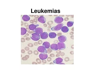
Chronic leukemias. CML CLL MDS. Chronic myeloid leuk. (CML). Comprises less than 20% of all leuk. & is seen most frequently in middle age .In more than 95% of patients there is replacement of normal Bone Marrow (BM) by cells with abnormal chromosome (Philadelphia or Ph chromosome).

E N D
Chronic leukemias CML CLL MDS
Chronic myeloid leuk. (CML) Comprises less than 20% of all leuk. & is seen most frequently in middle age .In more than 95% of patients there is replacement of normal Bone Marrow (BM) by cells with abnormal chromosome (Philadelphia or Ph chromosome)
Philadelphia chromosome: is abnormal chromosome 22 due to translocation of part of long arm (q) of chromosome 22 to another chromosome ,usually 9 , with translocation of part of chromosome 9 including ABL oncogene to chromosome 22 It is aquired abnormality of haemopoietic stem cells that present in all dividing granulocytes, erythroid & megakaryocytic cells also in some B & minority of T lymphocytes .
Clinical features: This disease occure in either sex M:F= 1.4:1 Moat frequently between the age of 40-60y .It may occurs in children & neonates & very old There is no predisposing factor but the incidence was increased in Japan after exposure to atomic bomb. 1-Symptoms of hypermetabolism e.g. weight loss, lassitude, anorexia or night sweat. 2- Splenomegaly is nearly always present & is frequently massive.
Clinical features 3- Features of anemia 4- Bruising, epistaxis, menorrhagia due to abnormal platelet function. 5- Gout or renal impairment due to hypercalcaemia from excessive purine breakdown 6- Rare symptoms include visual disturbances & priapism.
Lab. Findings: 1- Leucocytosis: is usually >50×109/L & some times >500×109/L . A complete spectrum of myeloid cells is seen in peripheral blood, the level of neutrophils & myelocytes exceed those of blast & promyelocytes 2- Ph chromosome on cytogenic analysis of blood or BM 3- BM is hyper cellular with granulopoietic predominance.
Lab. Findings: 4- Neutrophil alkaline phosphatase score is low 5- Increased circulating basophiles 6- Normochromic normocytic anemia 7- Platelets count may be increased, normal or decreased 8- Serum B12 & B12 binding capacity increased 9- Serum uric acid increased
Chronic lymphocytic leuk.(CLL) Account for 25% or more of leuk. seen in clinical practice & occurs chiefly in the elderly. The accumulation of large numbers of lymphocytes to 50-100 times the normal lymphoid mass in the blood, BM, spleen, lymph nodes & liver. The cells are monoclonal population of B- lymphocytes. T- cell chronic leuk. is uncommon.
Classification of chroniclymphoid leuk B-cell CLL 1-B-CLL 2-B-PLL 3-HCL 4-PCL T-cell CLL 1-T-CLL 2-T-PLL 3-Adult T- cell leuk./lymphoma.
Clinical features: 1- The disease occurs in older subjects & is rare before 40 y The M: F= 2:1 2- Symmetrical enlargement of superficial lymph nodes, the nodes are usually discrete & non tender. 3-Features of anemia may be present 4- Splenomegaly &hepatomegaly are usual in late stages 5- Bacterial or fungal infections are common in late stages
Clinical features 6- Patient with thrombocytopenia may show bruising or purpura. 7- Excessive reaction to vaccination & insect bite may occur 8- Skin infiltration is present in small number of patients 9-Tonsillar enlargement may be a feature 10- Many cases are diagnosed when routine blood test is performed (accidentally)
Lab. Findings 1- lymphocytosis : the absolute lymphocyte count is>5×10⁹/L & may be up to 300 or more . between 70-99% of white cells in blood film appear as small lymphocytes .Smudged or smear cells are also present.
2- Normochromic ,normocytic anemia is present in late stages due to: BM infiltration Hypersplenism Autoimmune hemolytic anemia
Lab. Findings 3- Thrombocytopenia occurs in many patients 4- BM aspirate shows lymphocytic replacement of normal marrow elements. Lymphocytes comprise 25-95% of all cells 5-reduced concentration of S.Ig & this becomes more marked with advanced disease. 6-Trisomy12 or t (11:14) are the most frequent chromosome findings
Course & prognosis.CLL divided into 5 stages according to Rai classification
These stages correlate with different prognosis e.g. Rai 0 with mean survival of 12 y Rai 1 with mean survival of 8 y
Myelodysplastic syndromes (MDS) A large group of acquired neoplastic disorders of BM , most common in elderly & characterized by increased BM failure with quantitative & qualitative abnormalities of all 3 myeloid cell lines (red cells, granulocyte/monocyte &platelets ) due to defect of stem cells & ineffective haemopoiesis so that cytopenias often accompany a marrow of normal or increased cellularity. In most cases , the disease arises de novo , but in significant proportion chemotherapy &/or radiotherapy has previously been given for another hematological diseases.
Clinical features 50% of patients are >70 years, &<25% are <50 y old. -Male are more commonly affected. - The evolution is often slow & the disease may be found by chance -The symptoms if present are that of anemia, infections or of easy bruising or bleeding.
Clinical features -In some patients, transfusion dependant an. dominates the course. While in others recurrent infection or spontaneous bruising or bleeding is the major clinical problem. -Neutophils, monocytes & platelets are often functionally impaired lead to spontaneous infections, bruising or bleeding regardless the severity of cytopenias. -The spleen is not usually enlarged except in CMML in which gum hypertrophy & lymphadenopathy may occur.
Lab. Findings: 1- peripheral blood: pancytopenia is frequent finding The red cells are usually macrocytic or dimorphic but occationally hypochromic , normoblast may be present., the reticulocyte count is low . Granulocytes are often reduced &may show lack of granulation .Their chemotactic,phagocytic &adhesive function are impaired. The pelger abnormality (single or bilobed nucleus) is often present. In CMML, monocytes are>1×10⁹/L in the blood & total WBC >100×109/L . The platelets may be unduly large or small & are usually decreased in number but in 10% of cases are elevated. In poor prognosis cases myeloblasts are present in blood
2-BM: the cellularity is usually increased, ring sidroblast may occur in all 5 FAB types, but by definition they are>15% in RARS Multinucleated normoblasts & other dyserythropoietic features are seen The granulocyte precursors show defective primary & secondary granulation Megakaryocytes are abnormal with micto , small binuclear or polynuclear forms BM biopsy shows fibrosis in 10% of cases.
3-Chromosomal & oncogene abnormalities: Cytogenic abnormalities are more frequent in secondary than primary MDS & most commonly constitute partial or total loss of chromosome 5,7,Y or trisomy 8 RAS oncogene (N-RAS) mutation occur in 20% of cases FMS mutation occur in 15% of cases