Download
1 / 55
630 likes | 1.13k Views
Post partum Haemorrhage. Assistant Prof. Dr.Esraa AL- Maini. More than 100,000 maternal deaths worldwide result from obstetric hemorrhage each year American college of obs and Gyn 140.000 deaths per year or 1 women every 4 minutes. Primary postpartum hemorrhage (PPH)

E N D
Post partum Haemorrhage • Assistant Prof. Dr.Esraa AL-Maini
More than 100,000 maternal deaths worldwide result from obstetric hemorrhage each year American college of obs and Gyn 140.000 deaths per year or 1 women every 4 minutes
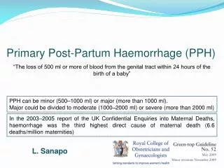
Primary postpartum hemorrhage (PPH) • The traditional definition of primary PPH is the loss of 500 ml or more of blood from the genital tract within 24 hours of the birth of a baby. • Secondary PPH is defined as abnormal or excessive bleeding from the birth canal between 24 hours and 12 weeks postnatal.
PPH can be classified into minor (500-1000 ml) or major (more than 1000 ml). • Major can be further subdivided into • Moderate (1001-2000 ml) • Severe (more than 2000 ml). • In women with lower body mass (e.g. less than 60 kg), a lower level of blood loss may be clinically significant
Risk factors for PPH Retained placenta Tissue Placenta accrete Tissue Episiotomy Trauma Perineal laceration Trauma General anaesthesia Tone The four Ts Multiple pregnancy Tone Previous PPH Tone Pre-eclampsia Thrombin Fetal macrosomia Tone Failure to progress in second stageTone Prolonged third stage of labour Tone
Causes of post partum haemorrhage (another busy slide) • Thrombin: abnormalities of coagulation • Pre-existing states Haemophilia A • Idiopathic thrombocytopenic purpura • von Willebrand's disease • History of previous PPH • Acquired in pregnancy Gestational thrombocytopenic • Pre-eclampsia with thrombocytopenia e.g. HELLP • Disseminated intravascular coagulation Gestational hypertensive disorder of pregnancy with adverse conditions • in utero fetal demise • severe infection ,abruption • amniotic fluid embolus Therapeutic anticoagulation Tone: abnormalities of uterine contraction Over distension of uterus Intra-amniotic infection Functional/anatomic distortion of uterus Uterine relaxants, e.g. magnesium and nifedipine Bladder distension Tissue: retained products of conception Retained cotyledon or succenturiate lobe Retained blood clots Trauma: genital tract injury Lacerations of the cervix, vagina or perineum Extensions, lacerations at caesarean section Uterine rupture Uterine inversion
Antenatal anaemia should be investigated and treated appropriately as this may reduce the morbidity associated with PPHfor pregnancy • -( if Hb 11 at first contact and 10.5 at 28 weeks) should be investigated • -
Iron supplementation considered if indicated. • -It is recommended that parenteral iron therapy should be considered antenatally for -women with iron deficiency anaemia who do not respond to oral iron. • -Antenatal anaemia (Hb less than 9 g/dl) and greater blood loss at delivery and postpartum Preparation for Postpartum Hemorrhage • Minimizing risk – reducing blood loss at delivery-Uterine massage is of no benefit in the prophylaxis of PPH.-Prophylactic uterotonics should be routinely offered in the management of the third stage of labour in all women as they reduce the risk of PPH..
B-Women at low risk for post partum hemorrhage GOOD NEWS B- Women at increased risk of PPH • delivering vaginally, oxytocin (10 iu by intramuscular injection) is the agent of choice for prophylaxis in the third stage of labour. Intramuscular oxytocin should be administered with the birth of the anterior shoulder, or immediately after the birth of the baby and before the cord is clamped and cut. A higher dose of oxytocin is unlikely to be beneficial. • For women delivering by caesarean section, oxytocin (5 iu by slow intravenous injection) should be used to encourage contraction of the uterus and to decrease blood loss. • Ergometrine-oxytocin versus oxytocin alone not superior only increase the adverse effects of nausea and vomiting, and elevation of blood pressure 5 fold • oxytocin is superior to misoprostol in the prevention of PPH. • Carbetocin is licensed in the UK specifically for the indication of prevention of PPH in the context of caesarean delivery. Good news WE HAVE THE TOOLS
GOOD NEWS B- Women at increased risk of PPH B-Women at increase risk for post partum haemorrhage • Ergometrine-oxytocin may be used in the absence of hypertension as it reduces the risk of minor PPH (500-1000 ml might be superior to syntocinon alone to prevent PPH). • Intravenous tranexamic acid(0.5-1.0g), inaddition to oxytocin, at caesarean section to reduce blood loss in women at increased risk of PPH. , but not vaginal birth. thromboembolic events should be considered . Good news WE HAVE THE TOOLS
Management of PPH Estimated blood loss • 1- The visual estimation of peripartum blood loss is inaccurate ,that clinical signs and symptoms should be included in the assessment of PPH. • In pregnancy, pulse and blood pressure are usually maintained in the normal range until blood loss exceeds 1000 ml; • tachycardia, tachypnoea and a slight recordable fall in systolic blood pressure occur with blood loss of 1000-1500 ml. A systolic blood pressure below 80 mmHg, associated with worsening tachycardia, tachypnoea and altered mental state, usually indicates a PPH in excess of 1500 ml.
A multidisciplinary team • If major PPH (blood loss of more than 1000 ml) • Ongoing bleeding • Clinical shock
Measures for minor PPH (blood loss 500-1000 ml) without clinical shock: What can done • intravenous access (one 14-gauge cannula) • urgent venepuncture (20 ml) for: • group and screen • full blood count • coagulation screen, including fibrinogen • pulse, respiratory rate and blood pressure recording every 15 minutes • commence warmed crystalloid infusion
Measures for major PPH • A and B - assess airway and breathing • C - evaluate circulation • position the patient flat • keep the woman warm using appropriate available measures • transfuse blood as soon as possible, if clinically required • until blood is available, infuse up to 3.5 l of warmed clear fluids • Hydroxyethylstarch should not be used. What can done
The main therapeutic goals of the management of massive blood loss as maintaining • Hb greater than 80 g/l • platelet count greater than 50 x 109/l • prothrombin time (PT) less than 1.5 times normal • activated partial thromboplastin time (APTT) less than 1.5 times normal • fibrinogen greater than 2 g/l. • -CMV-seronegativeproducts should be used to avoid transmission of CMV to the fetus • -The UK policy of universal leucocyte depletion substantially reduces the risk of CMV transmission, In an emergency, should be given to avoid delay • -CMV-negative blood or platelets are not needed for transfusion during delivery or in the postpartum period. • -The routine use of rFVIIa is not recommended in the management of major PPH unless as part of a clinical trial.-
Full protocol for monitoring and investigation in major PPH (blood loss greater than 1000 ml) and ongoing haemorrhage or clinical shock • immediate venepuncture (20 ml) for: • cross-match (4 units minimum) • full blood count • coagulation screen, including fibrinogen • renal and liver function for baseline • continuous pulse, blood pressure recording and respiratory rate (using oximeter, electrocardiogram and automated blood pressure recording) • Foley catheter to monitor urine output • two peripheral cannulae, 14 gauge • consider arterial line monitoring (once appropriately experienced staff available for insertion) • consider transfer to intensive therapy unit once the bleeding is controlled or monitoring at high • dependency unit on delivery suite, if appropriate • documentation of fluid balance, blood, blood products and procedures
Clinicians should be prepared to use a combination of • pharmacological, mechanical and surgical • methods to arrest PPH. , a sequence of these measures should be instituted in turn until the bleeding stops.
Initial Steps for PPH • The simple mechanical and physiological measures of ‘rubbing up the fundus’ • Emptying the bladder to stimulate uterine contraction represent first-line management of PPH • Administer uterotonicagents • Examine lower genital tract for lacerations.
Medications for Uterine Atony METHERGINE “Speedy” OXYTOCIN “The Champ” Cytotec Inexpensive (?) Effective
oxytocin 5 iu by slow intravenous injection (may have repeat dose if bleeding continue) OXYTOCIN • . OXYTOCIN “The Champ”
METHERGINE • Ergometrine 0.5 mg by slow intravenous or IM • Ergometrine OR oxytocin as first-line agents for the treatment of PPH, • It seems appropriate to use both agents, although oxytocin is to be preferred initially, especially in women with hypertension or pre-eclampsia. METHERGINE “Speedy”
Prostaglandin F2 15-methyl • Carboprost 0.25 mg by intramuscular injection repeated at intervals of not less than 15 minutes to a maximum of eight doses (use with caution in women with asthma) • Misoprostol 800 micrograms. Misoprostol by any root took I.0-2.5 hours to increase uterine tone
In summary intravenous bolus of oxytocinshould be given slowly in a dose of not more than 5 iudose may be repeatedIf bleeding occurs at the time of caesarean section, intramyometrial injection of carboprost may be used (although not licensed). It is also possible to inject intramyometrialcarboprost through the abdominal wall in the absence of laparotomy. The recommended dose is 250 micrograms intramuscularly. This may be repeated every 15 minutes to a total dose of 2 mg (eight doses). However, if significant atonichaemorrhage continues after a third dose of carboprost, without significant improvement (i.e. 30 minutes or more after the first dose was given),
The patient should consider transfer to the operating theatre for that oxytocin infusion should be start oxytocininfusion (40 iu in 500 ml isotonic crystalloids at 125 ml/hour) unless fluid restriction is necessary Examination under anaesthesia, with an awareness of the impending need for laparotomy and/or hysterectomy. Surgical interventions should be initiated sooner rather than later.
Anesthesia While general anaesthesia with increased morbidity and mortality when compared with regional anaesthesia ,it may be preferable in patients who are haemodynamically unstable or who have a coagulopathy.
Surgical treatments • Intrauterine balloon tamponade • is an appropriate first-line 'surgical' intervention for control of a tonic PPHTamponadeusing various types of hydrostatic balloon catheter has superseded uterine packing • (packing not recommended for the control of atonicPPH) • Types • Foley catheter • Bakriballoon, • Sengstaken- Blakemore oesophageal catheter • Condom catheter • The urological Rusch catheter
Intrauterine balloon tamponade • Mechanism of action • -Exerting in inward-to-outward pressure > systemic arterial pressure: prevent continous bleeding. • Hydrostatic pressure effect of the balloon on the uterine arteries.
Positive test(Tamponade Test) • A ‘positive test’ (control of PPH following inflation of the balloon) indicates that laparotomy is not required, whereas a ‘negative test’ (continued PPH following inflation of the balloon) is an indication to proceed to laparotomy • Method : inserted into uterine cavity balloon inflated 75-150ml warm saline . • Warm saline speed up coagulation bleeding stop no further surgery needed • There is no clear evidence on how long the balloon tamponade should be left in place. In most cases, 4-6 hours of tamponade should be adequate to achieve and ideally it should be removed during daytime hours, in the presence of appropriate senior staff, in case further intervention is necessary. • papers have removed the balloon within 48 hours
Before its complete removal the blloon could be deflated but left in place to ensure that bleeding does not reoccur. Rate of deflation vary from 20 ml/hour to half the volume in the balloon at 12 hours.
Condom catheters • Urinary bladder was kept empty by indwelling Foley's catheter. • After putting the patient in the lithotomy position, the condom is inserted within the uterine cavity. • Inner end of the catheter remained within the condom. • Outer end of the catheter is connected with a saline set and the condom is inflated with 250-500 mL of running normal saline. Bleeding is observed, and when it is reduced considerably, further inflation is stopped and the outer end of the catheter is folded and tied with thread. • Oxytocin drip for at least 6 h after the procedure. • The uterine condom is kept tight in position by ribbon gauze pack or another inflated condom placed in the vagina. • The condom catheter is kept for 24-48 h and then is deflated gradually over (10-15 m) and removed. • Triple antibiotic coverage (amoxicillin [500 mg/6 h] + metronidazole [500 mg/8 h] + gentamicin [80 mg/8 hrs]) for 7 d.
Ruschballoonballoon has been described as preferable by its large capacity, ease of use and low cost successfully avoiding hysterectomy in (9I%) Rusch balloon and the condom catheter conforming naturally to the contour of the uterus do not allow drainage of the uterine cavity. Insufflation capacity of 1500 ml balloon was inflated with 400-500 ml of warm saline removed after 24 h following deflation at a rate of 20 ml/h
Balloon tamponade catheter Contours to uterine shape Dual lumen catheter that allows infusion of saline to expand the balloon while providing uterine drainage (provides drainage at the fundus)to monitor the progression of hemostasis
Multiple urinary Foley catheters inserted together applied to the oozing inner surface of the lower uterine segment The uterine incision site was then closed, and each of the balloonswas inflated with 35-75 cm3 of saline or water. Gentle traction was then applied to obtain a continuous tamponade effect, and the vagina was packed. •The catheters were then tied together, and plastic bag was used for the collection and measurement of blood loss prevent blood collection inside the uterine cavity and provide an accurate estimation of bleeding.
Sengstaken-Blakemore tube The Sengstaken-Blakemore two-balloon tube, originally designed for the management of bleeding oesophagealvarices, The distal, gastric balloon was filled with 300 ml of normal saline Subsequently, the proximal oesophageal balloon The greater cost of the Sengstaken-Blakemore tube in comparison to the Bakri balloon If insufflated to a large volume so urological Ruschballoon can be used
Bakri ‘SOS’ (Surgical Obstetric Silicone) balloon Capacity: up to 500 ml of saline Drainage channel: large bore Uses: PPH resulting from a low-lying placenta/placenta praevia Under US guidance, the balloon portion of the catheter is inserted into the uterus, making certain that the entire balloon is inserted past the cervical canal and internal ostium
Oxytocin infusion No evidence that an oxytocin infusion is obligatory for all causes of PPH. If the syntocinon is continued for the duration of balloon placement, this can range from 2 to 82 hours . Prolonged: hyponatraemia {cross-reactivity of the oxytocin with antidiuretic hormone receptors. Carbetocin, a synthetic analogue of oxytocin, with a half life of 4-10 times that of oxytocin is available. There were no significant changes in sodium, potassium or chloride values . Therefore, this may be a preferred drug in the presence of a uterine balloon for prolonged uterine contraction. Although not specifically mentioned, another means of increasing uterine tone is to encourage breastfeeding. However, this may be impractical or declined by the mother.
Antibiotic • to reduce the risk of iatrogenic infection • •E.g: cephalosporin. • •Duration: ± • prophylactic (single dose), • continued for 24-48 hours or • recommended for the duration of balloon usage Pain relief During insertion:Following a vaginal delivery: No anaesthetic analgesia (pethidine) may be used’. After insertion: no pain relief
Complications: • obstruction by uterine leiomyomata • inadvertent damage to the balloon during preparation of Sengstaken-Blakemore tube while cutting off the tip • • inability to place the balloon due to the presence of a B- Lynch • suture • insufficient insufflation requiring two balloons. • •air emboli if air is used as the distension medium for the balloon. • •uterine rupture from uterine overdistension • uterine perforation during insertion. • pregnancy reported following the use of the Rusch balloon • And following the use of a Bakri balloon in combination with a B-Lynch suture.
Haemostatic brace sutures Sutures material
B -LUNCH • The best known version, described by B-Lynch is • -particularly suitable when the uterus has already been opened for a caesarean section. • Requires hysterotomy for its insertion after vaginal delivery to ensure empty uterine cavity exclude abnormal placentation remove the large blood clots • . This may have a dual action of reducing uterine blood flow and compressing the bleeding surface • overall failure rate of sutures leading to hysterectomy was 25% • Risk factors for a hysterectomy included increasing age and vaginal delivery. In addition, a prolonged delay of 2-6 hours between delivery and uterine compression suture • There was no difference in failure rate among suture techniques.