Download
1 / 5
50 likes | 69 Views
Human VEGF ELISA Kit can be provided from Creative Diagnostics.<br><br>https://www.creative-diagnostics.com/Human-VEGF-ELISA-Kit-104214-463.htm<br>

E N D
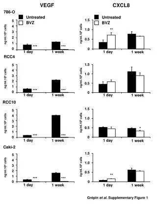
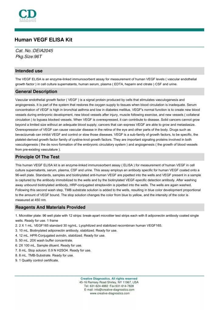
Human VEGF ELISA Kit Cat. No.:DEIA2045 Pkg.Size:96T Intended use The VEGF ELISA is an enzyme-linked immunosorbent assay for measurement of human VEGF levels ( vascular endothelial growth factor ) in cell culture supernatants, human serum, plasma ( EDTA, heparin and citrate ) CSF and urine. General Description Vascular endothelial growth factor ( VEGF ) is a signal protein produced by cells that stimulates vasculogenesis and angiogenesis. It is part of the system that restores the oxygen supply to tissues when blood circulation is inadequate. Serum concentration of VEGF is high in bronchial asthma and low in diabetes mellitus. VEGF's normal function is to create new blood vessels during embryonic development, new blood vessels after injury, muscle following exercise, and new vessels ( collateral circulation ) to bypass blocked vessels. When VEGF is overexpressed, it can contribute to disease. Solid cancers cannot grow beyond a limited size without an adequate blood supply; cancers that can express VEGF are able to grow and metastasize. Overexpression of VEGF can cause vascular disease in the retina of the eye and other parts of the body. Drugs such as bevacizumab can inhibit VEGF and control or slow those diseases. VEGF is a sub-family of growth factors, to be specific, the platelet-derived growth factor family of cystine-knot growth factors. They are important signaling proteins involved in both vasculogenesis ( the de novo formation of the embryonic circulatory system ) and angiogenesis ( the growth of blood vessels from pre-existing vasculature ). Principle Of The Test This human VEGF ELISA kit is an enzyme-linked immunosorbent assay ( ELISA ) for measurement of human VEGF in cell culture supernatants, serum, plasma, CSF and urine. This assay employs an antibody specific for human VEGF coated onto a 96-well plate. Standards, samples and biotinylated anti-human VEGF are pipetted into the wells and VEGF present in a sample is captured by the antibody immobilized to the wells and by the biotinylated VEGF-specific detection antibody. After washing away unbound biotinylated antibody, HRP-conjugated streptavidin is pipetted into the wells. The wells are again washed. Following this second wash step, TMB substrate solution is added to the wells, resulting in blue color development proportional to the amount of VEGF bound. The stop solution changes the color from blue to yellow, and the intensity of the color is measured at 450 nm. Reagents And Materials Provided 1. Microtiter plate: 96 well plate with 12 strips: break-apart microtiter test strips each with 8 adiponectin antibody coated single wells. Ready for use. 1 frame 2. 2 X 1 mL. VEGF165 standard 30 ng/mL. Lyophilized and stabilized recombinan human VEGF165. 3. 10 mL. Biotinylated adiponectin antibody, stabilized. Ready for use. 4. 12 mL. HPR-Conjugated avindin, stabilized. Ready for use. 5. 50 mL. 20X wash buffer concentrate. 6. 2X 100 mL. Sample diluent. Ready for use. 7. 8 mL. Stop soluion: 0.9 N H2SO4. Ready for use. 8. 8 mL. TMB-Substrate. Ready for use. 9. 1 Quality control certificate. Creative Diagnostics. All rights reserved 45-16 Ramsey Road Shirley, NY 11967, USA Tel: 631-624-4882 ·Fax:631-614-7828 E-mail: info@creative-diagnostics.com www.creative-diagnostics.com
Materials Required But Not Supplied 1. Microplate reader capable of measuring absorbance at 450 nm. 2. Precision pipettes ( 2 µL to 1 mL volumes ). 3. Multi-channel pipette ( 25 µL to 350 µL ). 4. Adjustable 1-25 mL pipettes for reagent preparation. 5. 100 mL and 1 liter graduated cylinders. 6. Absorbent paper. 7. Distilled or de-ionized water. 8. Log-log graph paper or computer and software for ELISA data analysis. 9. Tubes to prepare standard or sample dilutions. 10. Timer Storage Store all contents at 2 to 8 ℃. Specimen Collection And Handling Serum, EDTA, heparin or citrate anti-coagulated plasmas, cerebrospinal fluid, cell culture supernatants and urine are suitable sample types for use in the assay ( caution: separate plasma/serum and blood cells within 4 hours after collection, non- separated samples must be kept in temperatures from +2-8 ℃ ). Do not use grossly haemolyzed or lipemic specimens. If samples are to be run within 24 hours, they may be stored at +2-8 ℃; otherwise samples should be stored frozen ( at least between -18 to -32 ℃, but preferably < -70 ℃ ). Up to 3 freeze-thaw cycles have no effect on the VEGF levels of samples. Nonetheless, excessive freeze-thaw cycles should be avoided. Prior to the assay, frozen samples should be thawed as quickly as possible in tap water ( 18-25 ℃ ). Do not use 37 ℃ or 56 ℃ water bath for this purpose. Reagent Preparation Note: All reagents and samples must be allowed to equilibrate to room temperature ( 18-25 ℃ ) before use. 1. Antibody coated plate: Before opening the foil pouch, determine the number of strips required to test the desired number of samples, plus 16 wells needed for running standards and blanks in duplicate. Remove non-used strips from the plate-frame and return them to the foil pouch containing the desiccant for up to 3 months at 2-8 ℃. 2. Dilution of test standard: Dissolve the lyophilised VEGF standard with Sample Diluent volume shown on the label. VEGF standard is unstable after dissolving. Use immediately or keep on ice if not used within 1 hour after dissolving. 1 ). Take 100 µL of VEGF165 from kit standard tube containing 4200 pg/mL of VEGF and pipet into Standard tube 1. Add 500 µL of sample diluent to obtain VEGF concentration of 700 pg/mL in the first dilution tube ( total volume 600 µL ). 2 ). Add 200 µL of Sample Diluent to all other 4 dilution tubes. Take 100 µL from the first tube ( 700 pg/mL ) and start 3-fold serial dilutions in dilution tubes as described in the figure by mixing several times with the pipette in each tube ( Total of 5 dilution tubes ). 3 ). 200 µL of Sample Diluent serves as zero standard ( 0 ng/mL ) in tube 6. 3. Sample preparation and dilution: Dilution of samples is not required for initial screening. Samples that exceed the measuring range should be diluted serially in sample diluent and measured again. Samples with absorbance values >1.900 can be serially diluted 1:2, 1:4, 1:8, or further if necessary. The dilution factor must be taken in account when calculating the results. Dilute and store all samples in tubes or plates made of material with low binding surface, such as polypropylene. 4. Preparation of reagents: 1 ). Wash Buffer: If the 20x concentrated Wash Buffer contains visible crystals, warm it at 37 ℃ and mix gently until dissolved. Dilute 1:20 with de-ionized or distilled water ( e.g. 25 mL of Wash Buffer Concentrate and 475 mL distilled water to yield 500 mL of 1x Wash Buffer ). Check the pH of the diluted wash buffer and adjust to 7.4 if necessary. Creative Diagnostics. All rights reserved 45-16 Ramsey Road Shirley, NY 11967, USA Tel: 631-624-4882 ·Fax:631-614-7828 E-mail: info@creative-diagnostics.com www.creative-diagnostics.com
2 ). Vortex mix green colored biotinylated antibody solution gently before use. 3 ). Vortex mix blue colored peroxidase ( HRP ) labeled avidin gently before use. Assay Steps 1. Bring all reagents and samples to room temperature ( 18 - 25 ℃ ) before use. It is recommended that all standards and samples are run at least in duplicate. Leave some wells as a reagent blank ( 2 to 4 wells ). 2. Pipette 50 µL of sample and 50 µL of each diluted standard starting from 1200 pg/mL into appropriate wells. 3. Pipette 50 µL of sample diluent to the wells which will be used as a blank. 4. Incubate 1 hour at room temperature Wash 5x with 1x Wash Solution ( 300 µL each ) 5. To wash manually: Empty plate contents. Use a multi-channel pipette to fill each well with 300 µL of diluted wash buffer, then empty plate contents again. Repeat procedure 4 additional times for a total of FIVE washes. Gently blot plate onto paper towels or other absorbent material. Never let reaction wells dry. Continue to the next step without delay or interruption. 6. For automated washing: Aspirate all wells and wash 5 times with 300 µL diluted wash buffer. Blot plate onto paper towels or other absorbent material. Never let reaction wells dry. Continue to the next step without delay or interruption. 7. Promptly add 50 µL of green colored Biotinylated VEGF detection antibody to all wells 8. Tap the plate gently by hand to homogenize your mixture. Avoid touching to the reaction wells with pipet tip. 9. Incubate at room temperature for 1 hour without shaking. 10. Wash 5 times 5x as described in Step 3. 11. Add 50 µL of prepared HRP-conjugated avidin solution ( ready to use ) to each well. 12. Incubate for 30 minutes at room temperature. 13. Wash 5 times as described in Step 3 14. Using a multichannel pipet, promptly add 50 µL of TMB ready to use substrate reagent to each well. 15. Incubate for 20 minutes at room temperature in the dark. 16. Add 25 µL of Stop Solution to each well. Read at 450 nm within 15 minutes. 17. Calculate the mean of reagent blank absorbance values and subtract it from all test well values ( standard and test samples ). Mean reagent blank absorbance value at 450 nm should be less than 0.200. 18. Calculate your results against standard curve, as outlined below. Quality Control 1. When not in use, kit components should be refrigerated. All reagents should be warmed to room temperature before use. 2. Microtiter plates should be allowed to come to room temperature before opening the aluminum pouches. 3. Once the desired number of strips has been removed, immediately reseal the pouch and store at 2 - 8 ℃ to maintain plate integrity. Protect from humidity. 4. Samples should be collected in pyrogen/endotoxin-free tubes. 5. Samples should be frozen if not analyzed shortly after collection. Avoid multiple freeze-thaw cycles of frozen samples. Thaw completely and mix well prior to analysis. 6. When possible, avoid use of badly hemolyzed or lipemic sera. If large amounts of particulate matter are present, centrifuge or filter prior to analysis. 7. It is recommended that all standards, controls and samples be run in duplicate. 8. Samples that contain > 15 ng/mL Adiponectin should be diluted serially with Sample Diluent. 9. When pipetting reagents, maintain a consistent order of addition from well-to-well. This ensures equal incubation times for all wells. 10. Cover or cap all reagents when not in use. 11. Do not use reagents after the kit expiration date. 12. Read absorbances within 15 minutes of assay completion. 13. In-house controls should be run with every assay. Creative Diagnostics. All rights reserved 45-16 Ramsey Road Shirley, NY 11967, USA Tel: 631-624-4882 ·Fax:631-614-7828 E-mail: info@creative-diagnostics.com www.creative-diagnostics.com
14. All residual wash liquid must be drained from the wells by efficient aspiration or by decantation followed by tapping the plate forcefully on absorbent paper. Never insert absorbent paper directly into the wells. 15. Because TMB Chromogen is light sensitive, avoid prolonged exposure to light. Also avoid contact between Stabilized Chromogen and metal, or color may develop. Calculation The standard curve must be determined individually for each experiment. Correct the absorbance values of all standards by subtracting from them the O.D. value of the reagent blank ( Bl = only sample diluent ). Calculate the mean absorbance value for each standard from the duplicates. The standard curve is used to determine the amount of VEGF in an unknown sample. The standard curve is generated by plotting the average O.D. ( 450 nm ) obtained for each of the standard concentrations on the vertical ( Y ) axis versus the corresponding VEGF concentration ( pg/mL ) on the horizontal ( X ) axis. Construct the standard curve using graph paper or statistical software. If samples generate values higher than the highest standard, dilute the samples with sample diluent and repeat the assay. Note that the concentration read from the standard curve must be multiplied by the dilution factor. Typical Standard Curve The following standard curve is obtained for the various VEGF standards over the range of 0 to 1200 pg/mL. The graph also shows the calibration data of WHO reference reagent compared to the kit standard. Detection Range 9-700 pg/mL Sensitivity <5 pg/mL Creative Diagnostics. All rights reserved 45-16 Ramsey Road Shirley, NY 11967, USA Tel: 631-624-4882 ·Fax:631-614-7828 E-mail: info@creative-diagnostics.com www.creative-diagnostics.com
Specificity Recongnize both natural and recombinant human AEGF165. Isoform VEGF121 cross-reacts 100 % in the assay. No cross- reactivity was observed with the following recombinathuman proteins: IL-1, IL-1ß, IL-2, IL-3, IL-4, IL-5, IL-6, IL-7, IL-8, IL-9, IL- 10, IL-12, IL-13, TNF α-, TARC Reproducibility Intra-Assay Precision: ≤ 6% Inter-Assay Precision: ≤ 10% Inter-Lot Precision: ≤ 12% REFERENCES 1. Ferrara, N., Houck, K., Jakeman, L., and Leung, D.W. ( 1992 ). Molecular and biological properties of the vascular endothelial growth factor family of proteins. Endocr. Rev. 13, 18-32. 2. Ferrara N. ( 2002 ). Timeline: VEGF and the quest for tumour angiogenesis factors. Nat Rev Cancer 2, 795-803. 3. Tischer, E., Mitchell, R., Hartmann, T., Silva, M., Gospodarowicz, D., Fiddes, J.C., and Abraham, J.A. ( 1991 ). The human gene for vascular endothelial growth factor. Multiple protein forms are encoded through alternative exon splicing. J. Biol. Chem. 266, 11947-11954. Creative Diagnostics. All rights reserved 45-16 Ramsey Road Shirley, NY 11967, USA Tel: 631-624-4882 ·Fax:631-614-7828 E-mail: info@creative-diagnostics.com www.creative-diagnostics.com