Download
1 / 23
250 likes | 405 Views
Molecular Imaging Lecture 2. PET/CT NUCLEAR IMAGING BY Dr. H. Hawesa hhawesa@ksu.edu.sa. Molecular Imaging.

E N D
Molecular ImagingLecture 2 PET/CT NUCLEAR IMAGING BY Dr. H. Hawesa hhawesa@ksu.edu.sa
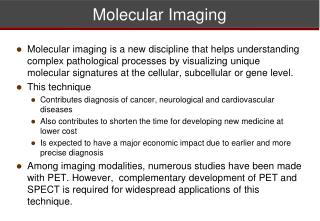
Molecular Imaging • Def: “ is a new biomedical research discipline enabling the visualization , characterization, and quantification of biologic process taking place at cellular and subcellular level within intact living subjects including patients”. • Molecular imaging Includes: PET, US, MRI, CT
Positron Emission Tomography • A technique involves detection of two gamma photons (each 115 KeV). • Positron emitting radionuclides are cyclotron produced (located close to PET scanner). • Fluorine-18 [T½=110 min] is commonly used (<2 hours). • PET allows high quality, quantitative Imaging.
Availability of radiotracers through commercial distribution network. • Mobile PET Imaging services. • Payment approved by medical insurance companies. • New generation of dual imaging modality (PET/CT). • Currently>1,000 PET/CT scanners are being sold worldwide than PET alone. • One PET/CT scanner for each 1 million population.
Difference between PET & SPECT • No need for conventional collimation (electronic). (PET has better sensitivity due to electronic collimation). • Attenuating path through patient is independent of the exact location of the annihilation event.
Basic Physics of PET Positron Decay & annihilation • P→ n + β⁺ + v • Β⁺ emitted from the nucleus . Its travels a short distance, losing energy. • Β⁺ interact with e. • Both β⁺ + e are annihilated. • 2 gamma photons are produced. • 511 KeV (~ 180 ̊).
Choice of PET Detector Characteristics of PET detectors: • High stopping power [High] -( efficiency of the detector to absorb the total energy of 511 KeV). • Higher light output (better energy resolution). -light output per KeV of photon energy. • Decay time of light -(short decay time = higher efficiency of the detector).
PET scintillation crystal • BGO = Bismuth Germanate • LSO = Lutetium Oxyorthosilicate • Na(TI) = Sodium Iodide • GSO = Gadolinium Oxyorthosilicate • YSO = yttrium Oxyorthosilicate (not used in PET technology) • BaF₂ = Barium Fluoride ( shortest decay time, rarely used)
Detectors • Axial FOV is defined by width of the array of the rings • No. of rings 18-32 (depend on manufacturer). • No. of detectors per ring ranges in thousands • More no. of detectors/ring →More PMTs (better spatial resolution) • Width of detector elements (small element, better resolution) [3-5 mm in modern PET).
Typical Configuration of PET Detector • 8×8 elements (64 elements)connected to 4 PMTs • 4×4 elements (16 elements/ 1 PMT) • No. of crystals ( 9,000-18,000) • Spatial resolution (5-7 mm)
Coincidence events (T,S,R) • True (T), Scattered (S), Random (R)
Acquisition mode • 2D: (+ collimators) • Detectors separated by lead septa (collimator) • Sensitivity to true events is decreased • Scattered events is reduced. • 3D: (- collimators) • Without collimator or septa • Sensitivity to true events ↑ by 5 times • No. of Random events increased • Scattered events are increased • Require 3D reconstruction algorithm
Performance Parameters of PET Scanner • Performance parameters: • Resolution • Sensitivity • Noise • Contrast • Scattered radiation
Spatial resolution • Positron range (e.g. in water) (F-18 with Emax of 640 KeV, B range = < 1mm) Contribution = 0.2 mm on FWHM (Rb-82 with Emax of 3,350 KeV, B range = 10mm ) Contribution = 2.6 mm on FWHM • Noncolinearity of annihilation photon • Angle of noncolinearity = ± 0.25° Contribution = 1.5-2.0 mm on FWHM for 30-90 diameter • Small crystals grouped in block detector Contribution= ↓ resolution due to miss positioning of events in 4 PMTs
sensitivity • It’s a measure of counting efficiency of a PET scanner • Sensitivity depend on : • Geometric efficiency • Distance between the source & the detector • Diameter of the ring • Number of detectors in the ring • Detection efficiency (detector material, scintillation decay time) • LSO & GSO detectors are preferred to BGO ( better detector efficiency • PHA window • Dead time of the system • Sensitivity is ↑ at the center of FOV & gradually ↓ toward the periphery • As no. of rings ↑, sensitivity ↑ • In 3D mode , sensitivity ↑ by factor of 4to 8 compared to 2D mode [in 3D, scatter and random event ↑ significantly]
Noise • Image noise is the random variation in pixel counts across the image • Noise is given by [(1/√N)×100] N=total counts in pixel • Noise can be reduced by: • Acquiring data for a long time • Injecting more radiopharmaceutical • Improving the detection efficiency of the PET scanner
Contrast • It’s a measure of the delectability of an abnormality relative to normal tissue • Factors affect the contrast: • Count Density • Scattered radiation • Size of the lesion • Patient motion
Scatter Fraction (SF) • Scatter contribution ↑ with: - density & depth of the body tissue - window width • Scattered radiation ↑ the background to the image, thus ↓ the image contrast • In 2D mode, septa removes scattered events • SF is a parameter used to check the PET performance • SF = Cs / CpCs=scattered counts rate, Cp=prompt counts rate • The lower the SF, the better the image quality.
Summary Thanks