Download
1 / 22
320 likes | 845 Views
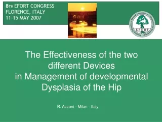
DEVELOPMENTAL DYSPLASIA OF THE HIP. Melih Güven, M.D Assoc. Prof. Yeditepe University Hospital Department of Orthopaedics and Traumatology Istanbul. Learning Objectives.

E N D
DEVELOPMENTAL DYSPLASIA OF THE HIP Melih Güven, M.D Assoc. Prof. Yeditepe University Hospital Department of Orthopaedics and Traumatology Istanbul
Learning Objectives 1. Should be able to define the term developmental dysplasia of the hip (DDH), and also explain the etiology and epidemiology. 2. Should be able to list the risk factors of DDH. 3. Should be able to explain the soft tissue and bone pathologies at the hip joint due to DDH 4. Should be able to define the basic examination methods for DDH for different ages 5. Should be able to define the treatment algorithms at different ages, to define prevention methods and also define the basic healthcare services for this reason
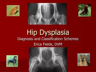
Description • Previously known as congenital dislocation of the hip implying a condition that existed at birth • Developmental encompasses embryonic, fetal and infantile periods • Includes congenital dislocation and developmental hip problems including subluxation, dislocation and dysplasia
Classification • Teratologic DDH • Typical DDH • Dysplasia • Subluxation • Dislocation
Incidence • Hip instability – 1% • True hip dislocation – 0.1% – 0.15% • Barlow stated that 60% stabilize in 1st week and 88% stabilize in first 2 months without treatment remaining 12% true dislocations and persist without treatment
Etiology – Risk factors high association with intrauterine molding abnormalities including metatarsus adductus and torticollis first born female baby ( 80% cases) left hip more common • Genetic and ethnic • increased native Americans but very low in southern Chinese and Africans • positive family history 12-33% • 10x risk if affected parent, 7X if sibling • intrauterine factors • breech position ( normal pop’n 2-4% , DDH 17-23% ) • oligohydroamnios • neuromuscular conditions like myelomeningocele
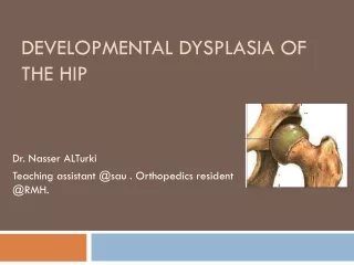
Pathologic Anatomy • Pathologic changes at soft tissues
Pathologic Anatomy • Pathologic changes at bone and joint
Diagnosis • Clinical risk factors • Physical exam • Ortolani Test • hip flexion and abduction , trochanter elevated and femoral head glides into acetabulum • Barlow Test • provocative test where hip flexed and adducted and head palpated to exit the acetabulum partially or completely over a rim
Diagnosis • Secondary adaptive changes occur • Limitation of abduction due to adductor longus shortening • Galleazi sign • flex both hips and one side shows apparent femoral shortening • Asymmetry gluteal, thigh or labial folds • Limb-length inequailty • Waddling gait and hyperlordosis in bilateral cases
Diagnosis Plain radiographs • Hip ultrasonography (USG) Alfa Beta
Natural History without Treatment • Barlow • 1 in 60 infants have instability ( positive Barlow) • 60% stabilize in 1st week • 88% stabilize in 2 months without treatment • 12 % become true dislocations and persist • Coleman • 23 hips < 3 months • 26% became dislocated • 13 % partial contact with acetabulum • 39% located but dysplastic feature • 22% normal • Because not possible to predict outcome all infants with instability should be treated
Goals of Treatment • Stable • Painless • Congruent • Dynamic and mobile Hip joints
Treatment Modalities • Conservative vs Surgical • Depends on • Type of DDH • Timing • Degree of the pathology • Previous treatments
Treatment Algorithm according to Age • 0 to 6 months • Goal is obtain reduction and maintain reduction to provide optimal environment for femoral head and acetabular development
Treatment Algorithm according to Age • 6 to 18 months • Surgery starts • Closed or open reduction and spica cast immobilization
Treatment Algorithm according to Age • Above 18 months • Open reduction is usually necessary • Additionally pelvic and femoral osteotomies
You should remember… • Risk factors of DDH • Definition of dysplasia, subluxation, dislocation • Soft tissue and bone pathologies at the hip joint due to DDH • Physical examination methods and imaging modalities for DDH • Treatment algorith of DDH