Download
1 / 29
330 likes | 483 Views
Endometriosis & Adenomyosis. Dr Ismaiel Abu Mahfouz. Endometriosis. Endometriosis. Definition Presence and growth of endometrial glands and stroma outside the endometrial cavity A benign condition Most cases of endometriosis Dx: 25 to 35 years Prevalence

E N D
Endometriosis & Adenomyosis Dr Ismaiel Abu Mahfouz
Endometriosis Definition • Presence and growth of endometrial glands and stroma outside the endometrial cavity • A benign condition Most cases of endometriosis Dx: 25 to 35 years Prevalence • Not known in the general population • 5-10% in Caucasian “Estimation” • 30- 40% in women with sub-fertility • 30% of women with chronic pelvic pain • 4-10% in women undergoing laparotomies
Why Endometriosis is important? Prevalent Distressing Associated with: Sub-fertility Chronic pelvic pain May invade adjacent organ; GIT. UT Typically affecting young women; 30s May present at extremes of reprod. age (rare) Scarification of old disease my cause obstructive symptoms in GIT and UT
Pathophysiology ?? Multifactorial Exact cause; not known Genetic predisposition Immune system Theories regarding pathogenesis The retrograde menstruation Endometrial tissue can grow in vivo and in vitro The müllerian metaplasia Metaplastic transformation of peritoneal mesothelium into endometrium The lymphatic spread 20% of women showed endometrial tissue in pelvic lymphatic system
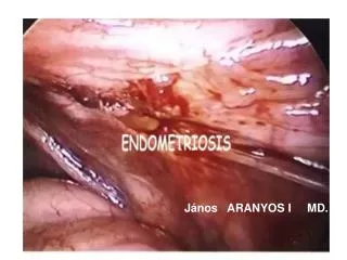
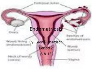
Endometriosis; sites The pelvis Ovaries (most common site, 70%) Broad and uterosacral ligaments Fallopian tubes Peritoneal surfaces of the POD Rectovaginal septum Vagina, vulva, appendix Extrapelvic site Laparotomy scars Lungs Forehead Axillae
Clinical presentation; Symptoms Risk factors: Caucasian. Nullip. High economic class Symptoms: Gynaecology symptom • Pain: Dysmenorrhea, dyspareunia, chronic pelvic pain • Pre and postmenstrual spotting • May cause heavy periods • Sub-fertility Extragenital symptoms • Dyschezia and PR bleeding • Haematuria • Masses in various places (Scar tissue) But: may be asymptomatic, and diagnosed during surgery
Clinical presentation; Signs Abdominal examination • Usually unremarkable, except severe disease • Tenderness following ruptured cyst • Mass Speculum • Bluish discoloration of cervix or vagina Pelvic examination • A small, tender nodule in POD or uterosacrals • Pelvic adnexal mass (tender, fixed) • Fixed ? Retroverted uterus / pelvic mass (adhesions) During Laparoscopy / laparotomy • Endometriotic spot • Endometrioma • Adhesions
Endometriosis; investigation Ultrasound scan • Adnexal mass • Endometrioma MRI • To investigate a deep endometriosis • Recto-vaginal masses
Endometriosis, DDx Pelvic inflammatory disease Acute salpingitis (? Hydrosaplinx) Haemorrhagic corpus luteum Benign ovarian cyst Malignant ovarian neoplasm Ectopic pregnancy
Endometriosis; Dx Suspected on the basis of clinical presentation Afebrile, pelvic pain; a firm, fixed, tender adnexal mass; and tender nodules in the POD Imaging Ultrasonic / MRI ( less frequently used) Adnexal mass; complex echogenicity, internal echoes suggestive of old blood Blood investigations (No Dx blood marker) CA-125 Frequently elevated PPV: 20% Should not be used to Dx endometriosis
Laparoscopy in endometriosis The definitive diagnosis Based on characteristic gross and histologic findings Failure to identify disease visually on laparoscopy or laparotomy is due to Older implants may have a very subtle appearance Deeper lesions may not be visible Therefore, biopsy of any suspicious lesions improves diagnostic accuracy Laparoscopy Diagnosis Staging Treatment
Endometriosis; staging The revised American Society for Reproductive Medicine classification of endometriosis Four stages: Minimal Mild Moderate Severe Based on the presence and size of the disease in various pelvic sites including: Peritonium Both ovaries Both tubes
Management Indications for treatment • Pain syndromes: Chronic pelvic pain, Dysmenorrhea Dyspareunia • Abnormal bleeding • Ovarian cysts • Sub-fertility caused by distortion of tubal and ovarian anatomy The choice of Rx depends on: • Age • Fertility plans • Severity of disease / symptoms • Site of disease • Involvement of other organ systems (GIT)
ManagementConservative, Medical, Surgical Conservative Pain killer Avoid hormonal Rx. in women trying to conceive Patient support groups Medical Aim: Toproduce atrophy of ectopic endometrium Agents: COCP, progestogens, GnRH agonist All equally effective in reducing pain Use limited by S.E Symptoms recurrence is common after Rx cessation
ManagementConservative, Medical, Surgical Surgical Laparoscopy Ablation of superficial lesions (laser or bipolar) Excision of nodules Endometrioma: De-roofed / excision Up to 70% of women report improvement 90%: Improvement persists for 1 year TAH+BSO: Final option
Endometriosis and sub-fertility 30-40% of patients with endometriosis have sub-fertility Pathophysiology of sub-fertility in endometriosis • Distortion of pelvic anatomy and tubal adhesions • Abnormal peritoneal and cellular function • Ovulatory and endocrine abnormalities • Impaired implantation Management of endometriosis in sub-fertility Hormonal medical Rx: • Not indicated • Causes anovulation, ? teratogenicity Surgical: • Of mild to moderate disease improves natural conception rates • Of sever disease: improve success at IVF
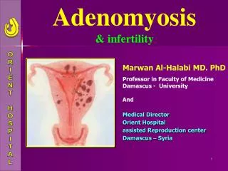
Adenomyosis Definition The extension of endometrial glands and stroma into the uterine musculature more than 2.5 mm beneath the basalis layer Adenomyosis Often is an incidental finding during a pathologic examination in up to 60% of women in their 40s About 15% have endometriosis Islands of adenomyosis do not participate in the proliferative and secretary cycles induced by the ovary
Adenomyosis; gross appearance The gross appearance of the uterus Diffuse enlargement of the uterus Thickened myometrium The endometrial cavity is enlarged Occasionally, may be confined to one part of the myometrium and of a round shape Adenomyoma vs fibroid on ultrasound: The distinction may not be clear Unlike fibroid: no distinct capsule between the adenomyoma and myometrium
Adenomyosis; Ultrasound Adenomyosis Fibroid
Clinical presentation Symptoms Many are asymptomatic Typically presentation Severe secondary dysmenorrhea HMB Occasional deep premenstrual dyparunia Pathophysiology of symptoms Adenomyosis islands do not respond to ovarian hormones But: Prostaglandin release and local inflammatory changes persist leading to pain and vasodilatation causing HMB
Clinical presentation Signs Abdomin: Normal Pelvic exam.: Symmetrically enlarged,“soft”, tender uterus in the premenstrual period Occasionally asymmetrical like a fibroid uterus Consistency of the uterus is softer than a fibroid Diagnosis: Usually on histology of hysterectomy Ultrasound scan ? MRI
Adenomyosis; Rx Depends on symptoms Conservative management NSAIDs Hormonal control COCP E2 patches DMPA Levonorgestrel IUD (Mirena IUS) Surgical; If conservative measures fail or contraindicated Hysterectomy Endometrial ablation “control the bleeding”