Download
1 / 27
360 likes | 629 Views
Schistosomiasis. Schistosoma haematobium Schistosoma mansoni Schistosoma japonicum. A Brief History. First described by German pathologist Theodore Maximilian Bilharz Bilharz performed autopsies on Egyptian patients who had died from the disease:

E N D
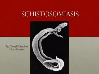
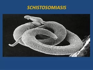
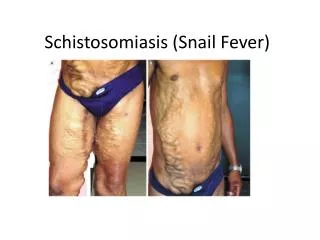
Schistosomiasis Schistosoma haematobium Schistosoma mansoni Schistosomajaponicum
A Brief History... • First described by German pathologist • Theodore Maximilian Bilharz • Bilharz performed autopsies on Egyptian patients who had died from the disease: found male & female parasite eggs in the liver portal system, bladder. • Later seen in Japan, called Katayama fever • Symptoms: rash on legs, fever, diarrhoea, bloody stools emaciation, edema death.
Schistosoma • General character: • Morphology • Reproduction system • Importance
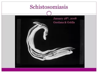
Morphology • Size: - Female 12 to 26 mm - Male 6 to 22 mm The three main species infecting humans are Schistosoma haematobium, S. japonicum , andS. mansoni. Two other species, more localized geographically, areS. mekongiandS. intercalatum
Adult an larve of Sch. Schistosomulum
Geographic Distribution • Schistosoma mansoniis found in parts of South America and the Caribbean, Africa, and the Middle East; • S. haematobiumin Africa and the Middle East; and • S. japonicumin the Far East. • Schistosoma mekongiandS. intercalatumare found focally in Southeast Asia and central West Africa, respectively.
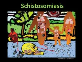
Schistosoma spp. cercariae are the infective forms. After encountering the skin, the cercariae penetrate and lose the tail transforming into schistosomulae
S. haematobium Biomphalaria
S. mansoni Oncomelania
Schistosoma egg • JK Sch.mansoni egg Sch. japonicum egg Sch. Haematobium egg
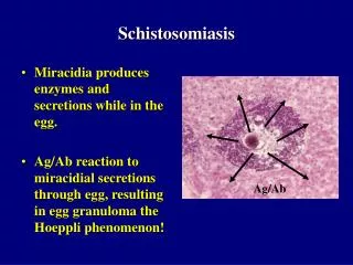
Laboratory Diagnosis • Microscopic identification of eggs in stool or urine is the most practical method for diagnosis. • Stool examination should be performed when infection withS. mansoniorS. japonicumis suspected, • and urine examination should be performed ifS. haematobiumis suspected. Tissue biopsy (rectal biopsy for all species and biopsy of the bladder forS. haematobium) may demonstrate eggs when stool or urine examinations are negative.