Download
1 / 23
250 likes | 296 Views
Left Laparoscopic Partial Nephrectomy. Louis R. Kavoussi M.D. Professor of Urology & Ardeshir R. Rastinehad D.O. Resident in Urology The Institute of Urology at the North Shore LIJ Health System. Patient Selection. Indications Enhancing renal mass Relative Contraindications

E N D
Left Laparoscopic Partial Nephrectomy Louis R. Kavoussi M.D. Professor of Urology & Ardeshir R. Rastinehad D.O. Resident in Urology The Institute of Urology at the North Shore LIJ Health System
Patient Selection • Indications • Enhancing renal mass • Relative Contraindications • Vena caval thrombus • Multiple prior abdominal surgeries • Significant medical comorbidities • Contraindications • Peritonitis • Uncorrected coagulopathy
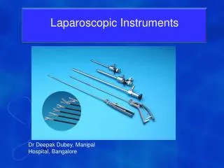
Equipment • PATIENT POSITIONING • Gel bolster, Gel pads • 3" Adhesive Silk tape • Pillow, Heel pads • Sequential compression device • Foley catheter, orogastric tube • ENERGY/IMAGING : • Monopolar and Bipolar • Argon Beam Coagulator (ABC) • Ligasure Atlas (10mm) [Valley lab] • Laparoscopic ultrasound probe VIDEO/CAMERA EQUIPMENT: • Laparoscopes (10 and 5 mm 30) • Thompson laparoscope holder • Hot water thermos • Video tower • HD monitors (2) • Camera unit • Light source • Insufflator • DVR and printer
SUTURES: Renorrhaphy stitches: O-Vicryl on CT needles (15 cm) Lapra-Ty Skin: 4-0 Caprosyn ADDITIONAL: Surgicel 10 mm Endoclip Lapra-Ty clips 15 Fr Round Blake drain and 100 cc reservoir Equipment TROCARS/INSTRUMENTS: • Trocars: 12 mm (3), 5 mm (2) • Major tray • Veress needle • Standard Laparoscopy Instruments • 10 mm laparoscopic scissors • Laparoscopic Bull Dog Clamps • Irrigator/aspirator • [Strykeflow irrigation system, Stryker]
Patient Positioning • OR table lined with gel pad • 30º modified flank, supported by gel pads/bolster • Lower legs supported by pillow – (slight knee flexion) • Left arm draped over chest, supported by gel pads • Arms, hips, and lower leg secured by tape over eggcrate padding and towel • Table tested to ensure patient secure with full “airplane” tilt
Patient Positioning Arm supported by gel pads; draped across abdomen and secured Pillow under knees with slight flexion; legs parallel
Gel roll placed behind scapula and back Secured at level of arm and waist
Orogastric tube and foley “Egg Crate” foam at all pressure points
Pneumoperitoneum/Trocars • Veress needle placed at umbilicus • 15 mm Hg CO2 pneumoperitoneum • Primary 12 mm camera port at umbilicus • (Laterally shift trocars in obese patients) • Second 12 mm trocar • Directly lateral to the camera port at midclavicular line • Accessory 5 mm trocar • Approximately 8 cm superior to the camera port
Trocar Placement Primary (12 mm) working ports 5 mm accessory port
Procedure Outline • Establish pneumoperitioneum and place the trocars • Mobilization of the colon • Identification of ureter • Dissection of renal hilum and upward retraction of ureter/lower pole • Enter Gerota’s fascia, identify the mass using intra-operative ultrasound • Hilar occlusion with bulldog clamps • Excise mass and place in entrapment bag • Renorrhaphy • Remove the bulldog clamps and the specimen
4. Dissection of renal hilum and upward retraction of ureter/lower pole
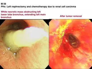
5. Enter Gerota’s fascia and identify the mass using intra-op ultrasound
9. Remove the bull dog clamps and the specimen (Clamp time = 26 minutes)
Technical points • Tenting the ureter anteriorly and “marching” cephalad facilitates rapid identification and dissection of the renal hilum • Intraoperative ultrasound must be available to identify and delineate renal mass, especially with endophytic lesions
Technical points • A mini-lap pad should be placed in the abdomen prior to releasing the bull dog clamps as it may be used to compress and tamponade unexpected bleeding from the renal bed • O-Vicryl on CT needles (15 cm), Lapra-Ty, and surgicel bolsters are used for hemostasis
Credits Surgeon: Louis R. Kavoussi M.D. Chairman of Urology North Shore LIJ – Health System New Hyde Park, NY Video editors: Ardeshir R. Rastinehad, Michael C. Ost, Lee Richstone Evan R. Eisenberg Institute of Urology North Shore LIJ – Health System