Download
1 / 11
110 likes | 265 Views
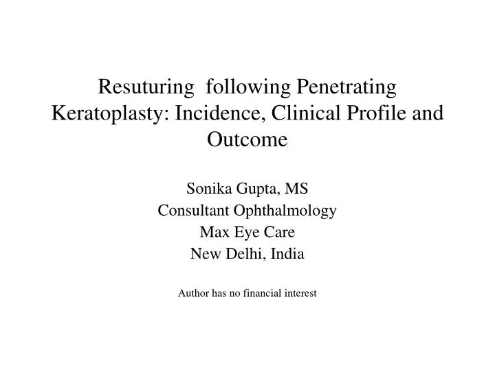
Resuturing following Penetrating Keratoplasty: Incidence, Clinical Profile and Outcome. Sonika Gupta, MS Consultant Ophthalmology Max Eye Care New Delhi, India Author has no financial interest. Purpose of study.

E N D
Resuturing following Penetrating Keratoplasty: Incidence, Clinical Profile and Outcome Sonika Gupta, MS Consultant Ophthalmology Max Eye Care New Delhi, India Author has no financial interest
Purpose of study To evaluate the clinical profile and outcome of cases requiring resuturing following penetrating keratoplasty (PKP).
Methods Study design and participants: In a retrospective case analysis of 258 consecutive PKP procedures performed from July 2004 to June 2008, medical records of patients who were admitted for resuturing of the corneal grafts were analyzed. .
Methods • Main parameters analyzed: Indications for PKP, time from PKP to resuturing, causes of resuturing, post-resuturing complications, visual outcome, and graft status. • Surgical technique: Similar method in all patients that involved a donor button oversized by 0.5 mm and placement of 16 interrupted sutures or 20 bite continuous running sutures
Clinical pictures of some cases of PKP requiring resuturing Fig.1: wound dehiscence inferiorly Fig.2 : loose suture at 2’o clock Fig.4: wound gape with infiltrates inferiorly Fig.3: unsatisfactory wound closure
Results • Resuturing was performed in 8.9% (23 eyes of 23 patients) . • Mean age of patients = 49.74 ± 16.529 years ; 14 males, 9 females • The incidence of resuturing was greater in cases operated for infective keratitis (16/113;14.1%) than for other indications (7/145; 4.8%, p=0.009 chi- square test
Results Indications for PKP in resutured grafts were infective keratitis in 16 eyes (69.5%), bullous keratopathy 4 eyes (17.3%), corneal scar 3 eyes (13%).
Results The main causes of resuturing: loose sutures in 12 eyes (52.1%) , unsatisfactory wound closure 6 eyes (26%), wound dehiscence 3 (13%) and broken sutures 2 (8.7%).[Fig 6]
Results • The median time between PKP and resuturing was 14 days (range 1-120 days). • Complications : graft infection(13%) and endophthalmitis (4.3%). • Visual acuity of ≥ 6/18 observed in 39.1% eyes over a mean follow-up period of 8.6 ± 4.20 months.
Conclusion • Resuturing of corneal wound after PKP is required for various suture-and wound-related complications including loose sutures and wound dehiscence. • Our results suggest that resuturing is most commonly required for PKP done for infective keratitis. The presence of severe ocular surface inflammation in these patients may contribute to suture related problems. Close monitoring is recommended in such cases.
Conclusion • Sutureless surgical procedures like Descemet’s stripping automated endothelial keratoplasty (DSAEK) may be preferred in cases requiring corneal transplantation for endothelial decompensation. • Deep anterior lamellar keratoplasty (DALK) may be encouraged in superficial and stromal corneal disease as risk of wound dehiscence is very low in DALK . • With new technologies such as femtosecond laser, superior mechanical stability of corneal wound is achieved, thereby reducing the risk of wound dehiscence.