Download
1 / 29
290 likes | 502 Views
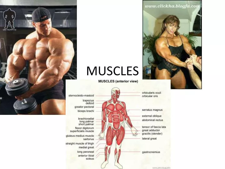
MUSCLES. 3 Types. Skeletal-striated/voluntary Smooth- involuntary Cardiac-heart, involuntary. Skeletal. Smooth. Cardiac. Skeletal Muscle Components. Skeletal muscle - numerous nuclei and mitochondria Fascia - dense CT, surrounds each muscle/separates

E N D
3 Types • Skeletal-striated/voluntary • Smooth- involuntary • Cardiac-heart, involuntary
Skeletal Muscle Components Skeletal muscle - numerous nuclei and mitochondria Fascia- dense CT, surrounds each muscle/separates - Tendon- cordlike, connects to bone - Aponeuroses- CT that connects bone - broad, fibrous sheets
CT • Epimysium – outermost layer, surrounds entire muscle • Perimysium– separated and surrounds the FASCICLES -(bundles) of muscle fibers • Endomysium– surrounds each individual muscle fiber • Many layers= ability to move independently, allows blood vessels and nerves to pass through
Sarcolemma- muscle fiber membrane • Sarcoplasm- inner fluid, cytoplasm • Myofibrils- indiv muscle fibers made of myofilaments within the sarcoplasm
MYOFILAMENTS. two types: MYOSIN – thick filaments, two twisted proteins strands, globular parts (cross bridges) ACTIN – thin filaments, double strands in helix
These filaments overlap to form dark and light bands on the muscle fiber • A band = dArk • thick (myosin) • I band = lIght • thIn (actin) • In the middle of each I band are Z lines. A sarcomere is one Z line to the other • arrangement of sarcomeres next to each other produces the STRIATIONS
Sarcoplasmic Reticulum–network of membranous channels surrounding myofibril • Transverse tubules (T tubules) – extend from sacroplasmic reticulum into sacrolemma. • Cristernae- 2 tubes surrounding T tubes
Muscles and Nervous System • Muscle contraction- movement of myofibrils: actin &myosin slide past one another, shortening sacromeres • Muscle fibers shortens - pulls attachments
Neuron- • Axon- extends / capable of conducting nerve impulse • Motor Neuron- control skeletal muscles
Synapse • Synapse- Space which info passes • Neurotransmitters- chemicals released into synapse • Neuromuscular junction- site where axon and muscle fiber meet- forms motor end plate • Nuceli/mitochondria abundant & sarcolemma is folded
Synaptic cleft- separates neuron membrane and membrane of muscle fiber
Steps for Contraction • Acetylcholine (Ach) –(neurotransmitter) released from end neuron • It diffuses across the gap to the muscle fiber • Muscle fiber is stimulated-impulse travels across fiber & into T tubules
4. Impulse reaches sarcoplasmic reticulum 5. Calcium ions are released 6. Cause linkages between actin and myosin 7. Actin filaments slide across the myosin 8. Muscle fiber shortens http://highered.mcgraw-hill.com/sites/0072437316/student_view0/chapter42/animations.html#
Sliding filament Theory The movement of the actin filaments over the myosin causes shortening of fiber
Fiber Relaxation 1. Cholinesterase is released 2. Inhibits acetylcholine 3. No muscle stimulation 4. Calcium ions reabsorbed into S.R 5. Actin returns to normal position 6. Muscle fiber relaxes