Download
1 / 1

E N D
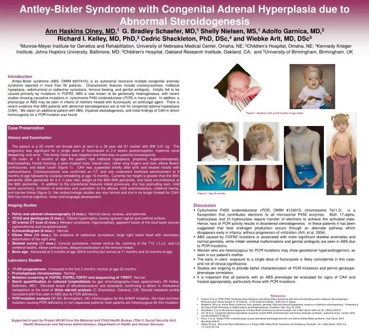
Antley-Bixler Syndrome with Congenital Adrenal Hyperplasia due to Abnormal SteroidogenesisAnn Haskins Olney, MD,1 G. Bradley Schaefer, MD,1 Shelly Nielsen, MS,1 Adolfo Garnica, MD,2Richard I. Kelley, MD, PhD,3 Cedric Shackleton, PhD, DSc,4 and Wiebke Arlt, MD, DSc51Munroe-Meyer Institute for Genetics and Rehabilitation, University of Nebraska Medical Center, Omaha, NE; 2Children’s Hospital, Omaha, NE; 3Kennedy Krieger Institute, Johns Hopkins University, Baltimore, MD; 4Children’s Hospital, Oakland Research Institute, Oakland, CA; and 5University of Birmingham, Birmingham, UK Introduction Antley-Bixler syndrome (ABS, OMIM #207410)) is an autosomal recessive multiple congenital anomaly syndrome reported in more than 50 patients. Characteristic features include craniosynostosis, midfacial hypoplasia, radiohumeral or radioulnar synostosis, femoral bowing, and genital ambiguity. Initially felt to be caused primarily by mutations in FGFR2, ABS is now known to be genetically heterogeneous, with recent studies showing causative mutations in cytochrome P450 oxidoreductase (POR) in many cases. In addition, a phenocopy of ABS may be seen in infants of mothers treated with fluconazole, an antifungal agent. There is recent evidence that ABS patients with abnormal steroidogenesis are at risk for congenital adrenal hyperplasia (CAH). We report an additional patient with ABS, impaired steroidogenesis, and initial findings of CAH in whom homozygosity for a POR mutation was found. Figure1: Newborn (left) and 8 months of age (right) • Case Presentation • History and Examination • The patient is a 25 month old female born at term to a 26 year old G1 mother with BW 3.01 kg. The pregnancy was significant for a single dose of fluconazole at 2-4 weeks postconception, maternal voice deepening, and acne. The family history was negative and there was no parental consanguinity. • On exam at 6 months of age the patient had midfacial hypoplasia, proptosis, trapezoidocephaly, brachycephaly, frontal bossing, a pear-shaped nose, lowset ears, rather long fingers and toes, elbow flexion contractures, and labial fusion (figure 1). CAH was suspected shortly after birth and treated initially with hydrocortisone. Craniosynostosis was confirmed on CT, and she underwent forehead advancement at 9 months of age followed by occipital remodeling at age 15 months. Currently her height is greater than the 95th percentile (50th percentile for a 3 ½ year old), weight at the 90th-95th percentile, and head circumference at the 98th percentile. In addition to the craniofacial features noted previously, she has protruding ears, mild facial asymmetry, limitation of extension and supination at the elbows, mild arachnodactyly, umbilical hernia, and narrow thorax (figure 2). Her endocrinologic studies are now normal and she is no longer treated for CAH. She has normal cognitive, motor and language development. • Imaging Studies • Pelvic and adrenal ultrasonography (3 mos.): Normal uterus, ovaries, and adrenals. • VCUG and genitogram (3 mos.): Clitoral hypertrophy, closely spaced vaginal and urethral orifices. • 3D cranial CT scan (6 mos.): Metopic synostosis, trigonocephaly, apparent fusion of the skull base sutures (sphenofrontal and occipitomastoid). • Echocardiogram (8 mos.): Normal. • Elbow films (10 mos.): No evidence of radioulnar synostosis; large right radial head with secondary deformation of the right ulna. • Skeletal survey (11 mos.): Coronal synostosis, narrow vertical ilia, notching of the T12, L1,L2, and L3 vertebral bodies, elbow contractures, delayed ossification of the femoral heads. • Bone age: Advanced at 3 months of age (BA 6 months) but normal at 11 months and 23 months of age. • Laboratory Studies • 17-OH progesterone: Increased in the first 2 months; normal at age 22 months. • Prometaphase chromosomes: Normal. • Mutation screeningin FGFR2, FGFR3, FGFR1 and sequencing of TWIST:Normal • Sterol quantificationin cultured lymphoblasts by gas chromatography-mass spectrometry (RI Kelley, Baltimore, MD): Elevated levels of dihydrolanosterol and lanosterol, confirming a defect in cholesterol metabolism at the level of Urine steroid analysis (C Shackleton, Oakland, CA): Steroid metabolic profile consistent with that seen in ABS due to POR deficiency. • POR mutation analysis (W Arlt, Birmingham, UK): Homozygous for the A284P mutation, the most common mutation causing POR deficiency in non-Japanese patients; both parents are heterozygous for this mutation. Figure 2: Age 25 months • Discussion • Cytochrome P450 oxidoreductase (POR, OMIM #124015, chromosome 7q11.2) is a flavoprotein that contributes electrons to all microsomal P450 enzymes. Both 17-alpha- hydroxylase and 21-hydroxylase require transfer of electrons to achieve the activated state. Hence, loss of POR activity results in disordered steroidogenesis. In these patients it has been suggested that fetal androgen production occurs through an alternate pathway which disappears early in infancy, without progression of virilization (Arlt, et al, 2004). • ABS caused by FGFR2 mutations is associated with more significant skeletal anomalies and normal genitalia, while milder skeletal malformations and genital ambiguity are seen in ABS due to POR mutations. • Women who are heterozygous for POR mutations may show gestational hyperandrogenism, as seen in our patient’s mother. • The early in utero exposure to a single dose of fluconazole is likely coincidental in this case, and not of clinical significance. • Studies are ongoing to provide better characterization of POR mutations and permit genotype- phenotype correlation. • It is important that all patients with an ABS phenotype be evaluated for signs of CAH and treated appropriately, particularly those with POR mutations. • References • Fukami, M et al. POR (P450 Oxidoreductase) Mutations and Antley-Bixler Syndrome with Abnormal Genitalia and/or Impaired Steroidogenesis: Molecular and Clinical Studies in 10 Patients. J Clin Endocrinol Metab. 2004 Oct 13 (Epub). • Braddock, SR et al. Antley-Bixler syndrome: face, skeleton, genitalia, CNS and adrenal hyperplasia; evidence of defective steroidogenesis. Presented at the David Smith Workshop on Malformations and Morphogenesis, Snowbird, Utah, Aug 18, 2004. • Shackleton, C et al. Biochemical diagnosis of Antley-Bixler syndrome by steroid analysis. Am J Med Genet. 2004 Jul 30; 128A(4):223-31. • Arlt, W et al. Congenital adrenal hyperplasia caused by mutant P450 oxidoreductase and human androgen synthesis: analytical study. Lancet. 2004 Jun 26;363(9427):2128-35. • Fluck, C et al. Mutant P450 oxidoreductase causes disordered steroidogenesis with and without Antley-Bixler syndrome. Nat Genet. 2004 Mar;36(3):228-30. • Kelley, RI et al. Abnormal Sterol Metabolism in a Patient With Antley-Bixler Syndrome and Ambiguous Genitalia. Am J Med Genet. 2002 Jun 15;110(2):95-102. Supported in part by Project #8188 from the Maternal and Child Health Bureau (Title V, Social Security Act), Health Resources and Services Administration, Department of Health and Human Services.