Download
1 / 27
290 likes | 1.01k Views
Biophysics of Heart and Circulation. Heart as a source of biopotentials. Heart as a pump. Blood flow in vessels. Ján Jakuš. Anatomy of Heart. - consists of 4 chambers: from 2 Atrias (A ) and 2 ven-tricles (V )

E N D
Biophysics of Heart and Circulation. Heart as a source of biopotentials. Heart as a pump. Blood flow in vessels. Ján Jakuš
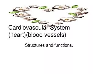
Anatomy of Heart - consists of 4 chambers: from 2 Atrias (A) and 2 ven-tricles (V) - wall of Left Ventricle (LV ) is 3 x thicker than a wall of the Right Ventricle (RV) - between RA and RV there is a tricuspid valve , bet-ween LA and LV is a bicuspid valve. - within the pulmonary artery, at a place where it leaves the heart, there is the pulmonary valve. - similarly, within the aorta there is the aortal valve - two coronary arteries bring the nutritiens and O2 for heart - Conductive system (nerve tissue within the heart): Sinoatrial node (SA), Atrio-ventricular node (AV ), Hiss bundle, Two branches of Tawara, Purkynie fibers.
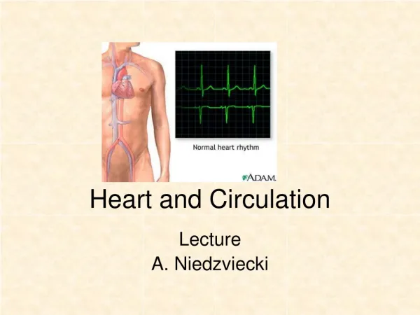
Spreading of Electric Depolarisation in the Heart ECG (see practicals) is a record of heart biopotentials from the body surface. Waves: P,T,U, Oscilations: Q,R,S,Segments: PQ,ST,QT, Intervals: PQ,ST,QT
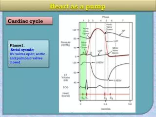
Dynamics of Heart Heart- 2 pumps, working together at the same time Systola - ejection of blood from ventricles- H.contraction Diastola - filling in ventricles with blood -H. relaxation Volume of blood from RV= Volume of blood from LV Systolic volume (SV)=70 ml ( volume of blood ejected from each ventricle during heart contraction) Diastolic volume of a ventricle (DV) = 130 ml (blood volume retained at ventricles during diastole Cardiac Output / min:CO= SV x Heart rate/ min. CO =70 x 72 = 5 (l/min.) Work of heart/60 years of life = move 60.000 kg of matter to the altitude of 8.000 m Work of LV=0,94 J/systole, Work of RV=0,19 J/ systole
Blood Pressures in Heart Right Atrium (systole/diastole) = +0,6 /-0,6 (kPa) Right Ventricle = 4,6 / 0 ! Pulmonary Artery = 4,6 / 1,2 Left Atrium = +0,6 /-0,6 Left ventricle = 16 / 0 ! Aorta = 16 / 10 At diastole, there are zero pressures within the ventricles ! At diastole, there are positive values of blood pressures both in the aorta and pulmonary artery, because ELASTICITY of the vessels (See practical –A model of elasticity)
Elasticity of aorta and arteries During ventricular systole - blood distends an aorta (kinetic energy -E- of a blood flow changes into potential E)-we can take Syst.BP Ventricular diastole-diameter of vessels decreases (potential Energy changes into kinetic one)- diastolic BP is measured Elasticity of vesselsenables: 1.Blood flow during diastole, 2.Diastolic BP, 3 Lower Work of heart
BLOOD CIRCULATION A./Big blood circuit- between LV and RA (there is a high BP and Pressure gradient PG =100 mm Hg (15kPa) Short blood circuit (lung circuit)-between RV and LA, PG =30mmHg (4 kPa) Portal blood circuit (btw. Hepatic artery and Portal vein) Fetal blood circuit –in foetus B./Distributive circuit(highly pressurized – aorta, arteries) Resistive circuit(arteriolies-contain smooth muscles in wall Diffusive circuit(capillaries-they create capillary loops) Capacitive circuit(veins and lymphatic vessels) Blood flows only along Presuure gradient ! Highest flow - in aorta- 30 cm/s, Lowest flow - in cap- illaries - 1mm/s
Blood flow Blood flowF ( l.s-1 )=Pressure of blood/ Resistance of vessel, F ( l.s-1 = P/ R • Blood flow depends linearly on Pressure Gradient, and non-linearly on an arteriolar resistance as well as on the composition of a blood (and thus on a blood viscosity) • Blood viscosity (is 4,5 x higher (4,5 mPa/s) then viscosity of distilled water. (e.g. when viscosity increases a blood flow decreases and vice versa. Poiseuille-Hagen Law Q= π . r4 . (P1-P2)(Q- amount of blood, r - vessel radius 8 . η . Lη - blood viscosity, L- lenght of vessel, • π- Ludolph number,P1,P2- B.Pressures)
Bernouli´s Law When blood flows through the narrow vessel the velocity of flow is higher but pressure of blood within is lower (and vice-versa)
TYPES OF BLOOD FLOWLaminar flow – parabolical shape of a streamline with max. velocity in the middle of the stream and the lowest velocity at the edges. Turbulent flow- velocity is very high, it creats soundRaynoldsove číslo R = ρ.d.v itinforms if flow is laminar(when R <, = 1100)η or turbulent ( when R > 1100)
Types of Blood Pressures(curve is taken from a direct measurement of BP)
Measurement of BP(non-direct method) Riva Rocci´s auscultatory method,Korotkov Sounds, Systolic and diastolic BP are taken (See practical for theory and procedure)
Filtration at Capillary Loop Capillary – a place for filtration of water and nutritiens and a resorptionof metabolic wastes , and for O2 a CO2 diffusion.
Failures of Cappilary Filtration and Resorption When the capillary transfer is damaged the OEDEMA (tissue swelling) appears. It is an accumulation of fluid among cells Causes: 1.Increase of Systemic Blood Pressure (hypertension), 2. Decrease of an Oncotic Presure- less than - 25 mmHg (a suction effect is lower and water retains within the tissue, that swells- as it appears in a disease Kwashiorkor, which is kind of protein deficiency) ( with typical big bellies in childrens) at economical poor countries 3.Increase in Capillary Permeability(something is wrong with the capillary wall- as seen in the Vibration disease or follo- wing some toxic effect of animal poisons on vessel permea- bility, e.g. snakes or scorpions ) 4. Disorders affecting the Lymphatic System ( the lymphatic circulation stops, because either cancer or parasite blocks the lymphatic vessels
TREATMENT:Angioplasty. Non-Surgical (Stenting) or Surgical (By-passing)
Thank You for Comming and Attention